Parts of the human brain (and functions)
The human brain has been described as the most complex system in the known universe, and not without reason.
It is made up of a network of glia, neurons and nerve pathways and is the most important part of the Central Nervous System, but its intricate structure and operation does not mean that we cannot make a classification of the main parts of the brain.
The main parts of the brain
In humans, The encephalon or brain is the part of the Central Nervous System that is located at the end of thespinal cord, inside the skull. It is, in short, the organ thanks to which we can perform the most complex mental operations and be aware, that is, a sense of self. It is precisely for this reason that within the brain there are a large number of structures working together to a large speed, a fact that makes the functioning of the brain, even today, a mystery in many of its aspects.
To begin to understand what we know about this complex machinery, it is essential to know the parts of the brain, that is, the way in which the structures that compose it can be classified. A good way to classify the different parts of the brain above can be taking into account the different formations that are formed within the head of a human embryo.
They are a total of three structures.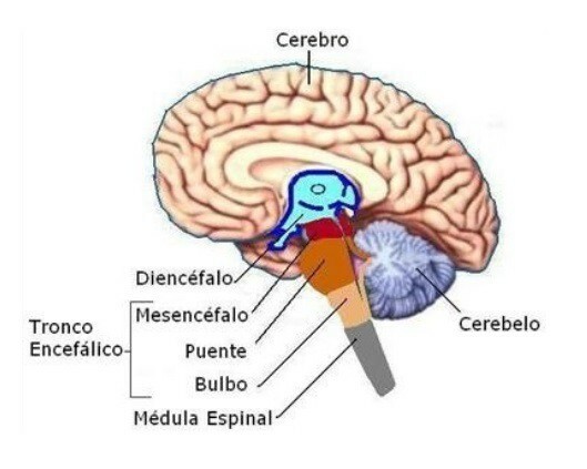
1.1. Rhombencephalon
It involves the upper part of the spinal cord and Throughout the development of the fetus, it will be transformed into the structures in charge of carrying out tasks essential for survival, such as heart rate and breathing control. It will end up transforming into the cerebellum, the brainstem bridge and the medulla oblongata, as we will see.
1.2. Midbrain
In human embryos it appears just above the rhombencephalon, and will gradually transform into the medial part of the brain, also in charge of performing a good part of the basic survival functions but it also acts as a bridge between the other two structures.
1.3. Forebrain
Located at the far end of the spinal cord and on the side closest to the face of the embryo, the forebrain is the formation that will be transformed into the parts of the brain that have appeared more recently in our evolutionary line and that, therefore, they have to do with the use of language, planning and finding creative solutions to new problems. As we will see, the two main structures to which the development of the rhombencephalon gives way are the diencephalon and the telencephalon.
The parts of the adult brain
Going into more detail, we can stop to see the different components of the brain in fully developed human beings. It is in this set of organs where we find all those parts of the brain that define the way our mind works.
Here we will see, first, the parts of the brain that are generated from the forebrain, and then move on to the midbrain area and the rhombencephalon, in that order.
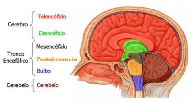
2.1. Telencephalon
The telencephalon is the part of the brain that is easiest to see with the naked eye, since it occupies most of the surface of the brain. Its components are the cerebral cortex, the basal ganglia and the limbic system..
2.1.1. Cerebral cortex
The cerebral cortex (or cortex) is the part of the brain that is rough and full of folds. It covers the rest of the brain above it, and is the area in which the information necessary to carry out the processes is integrated more complex mental structures, since the information that reaches this region has already been partially processed by other structures of the brain. The cortex is divided into two cerebral hemispheres that are almost symmetrical to the naked eye, although on a microscopic scale they are very different.
What's more, each hemisphere is made up of several lobes of the brain, each of which is more involved in certain mental processes. The lobes of the brain are these:
- Frontal lobe
- Parietal lobe
- Occipital lobe
- Temporal lobe
- Insula
- You can read more about it at this article on the brain lobes.
2.1.2. Basal ganglia
The second component of the telencephalon is the set formed by the basal ganglia. These are a group of structures located below the cerebral cortex and distributed symmetrically under each of the hemispheres. The basal ganglia are the globe pallidus, the putamen, and the caudate nucleus, which are complemented by a region known as the substantia nigra.
The basal ganglia are the parts of the brain that allow us to perform relatively complex and precise movements easily and almost automatically: write, speak, modify our facial expressions voluntarily, etc. Therefore, they semi-automatically monitor the way in which we carry out chains of movements that we have already practiced before many times until we mastered them, and at the same time allow us to learn them well, among others functions.
- To read more about this set of brain structures, you can visit the article dedicated to the basal ganglia.
2.1.3. Limbic system
The limbic system is a set of brain structures whose limits are quite diffuseas it mixes with many different parts of the brain. Its functions are related to the appearance and regulation of emotions and the bodily responses beyond the head that accompany them. That is why it is sometimes considered "the emotional brain" as opposed to the "emotional brain. rational "which would correspond to the areas occupied by the cerebral cortex (and especially the lobe frontal).
However, neither the limbic system nor the cortex can function well independently, and therefore this distinction between rational and emotional zones is very artificial, especially considering that we are not as rational as it might seem.
If you are interested in knowing more about this part of the brain, you can accessthis article on the limbic system.
2.1.4. Hippocampus
The hippocampus It is an elongated structure located in the inner part of the temporal lobes, one of the oldest regions of the cerebral cortex, present in the oldest forms of mammals. Its function is related to the storage and retrieval of memories, learning and spatial navigation.
- You can read more about this part of the brain at this article dedicated to the hippocampus.
2.1.5. Amygdala
The brain tonsil It is a set of neurons that are grouped on the inner face of the temporal lobe of each of the hemispheres. That is, like what happens with the hippocampus, it is one of those parts of the brain that is found in duplicate in each human brain, with one in each half (left and right) of the brain.
The brain amygdala is part of the limbic system, and it is one of the brain structures that are most important when it comes to relating emotional states with situations we live in; that is why it plays a key role in the mental processes related to emotional memory and the learnings related to it, which are very important. After all, knowing what emotions each type of stimulus or experience is paired with makes us adopt an attitude towards them and opt for some possible reactions and not others.
- You can read more about the amygdala at this article.
2.2. Diencephalon
The diencephalon is the second large structure that forms the forebrain, and is located just below the telencephalon, in the depths of the Central Nervous System. The parts of the brain that make up the diencephalon are basically the thalamus and the hypothalamus.
2.2.1. Thalamus
It is the largest part of the diencephalon, and it is the nucleus in which all the information that comes to us through the senses is integrated for the first time. (With the exception of smell, which reaches the brain directly through the olfactory bulb of each cerebral hemisphere). The thalamus sends this information to higher areas of the brain, so that the information that has begun to be synthesized in it continues to be processed there, and It is also capable of making it possible for the Autonomous Nervous System to react quickly to stimuli that may mean the presence of danger.
- To read more about this part of the brain you can read this article on the thalamus
2.2.2. Hypothalamus
The hypothalamus is located just below the thalamus, and is primarily responsible for making the entire body constantly in a state of homeostasis, that is, in balance in all senses: body temperature, blood hormone levels, breathing rate, etc.
In addition, thanks to its ability to make different glands of the body secrete hormones, it induces us to more or less high states of stress and general activation depending on what is happening in other parts of the brain. It is also the structure responsible for the appearance of the state of thirst and hunger.
- You can read more about the hypothalamus at this article
2.3. Brain stem
The brainstem, or brainstem, is the part of the brain that is most directly connected to the spinal cord, and it is also in charge of performing the basic tasks of maintaining vital functions such as involuntary breathing or heart rate. It is made up of the parts that evolve from the midbrain and the rhombencephalon. Its parts are as follows.
2.3.1. Midbrain
The midbrain is the part of the brain stem just below the diencephalon.. It is responsible for communicating the brain stem with higher structures and vice versa, and it also intervenes in the maintenance of automatic processes that allow us to survive. It is divided into the tectum and the tegmentum.
2.3.2. Boss
This structure is also known as the Varolio bridge or the brainstem bridge.. It is located just below the midbrain.
2.3.3. Medulla oblongata
It is the lower part of the brainstem, and its functions are very similar to those of the other two structures of this part of the brain. In addition, it is the link between the brain and the spinal cord. In the medulla oblongata is a part known as the decussation of the pyramids, which is where bundles of nerve fibers from the two hemifields (the left and right halves of the human body) intersect to pass from one side to the other; This explains why the right hemisphere is responsible for processing information from the left hand while the left is responsible for the other, for example.
- If you are interested in reading more about the brainstem, you can read this article
2.4. Cerebellum
Along with the medulla oblongata and pons, the cerebellum is the third major structure that evolves from the rhombencephalon. Also, the cerebellum and pons are part of a region called the metancephalon.
The cerebellum is one of the parts of the brain with a higher concentration of neurons and among its many functions the most studied is the regulation and monitoring of complex movements that require a certain coordination. It also has a role in maintaining balance when standing and walking.
- If you are interested in knowing more about the cerebellum, you can visit this article
Other related nervous system structures
The different parts of the brain not only work in coordination with each other, but they need the participation of other surgeons of the neuroendorine system.
These structures and systems, which do not belong to the brain itself, are the cerebral nerves (or cranial nerves) and the Autonomous Nervous System (ANS).
Cranial nerves
The cranial nerves are bundles of axons that emerge from different points in the lower brain and go to other parts of the body bypassing the spinal cord. This is what distinguishes them from the rest of the nerves, which do not come out from the different parts of the brain but from various sections of the spinal cord.
Examples of the cranial nerves are the trigeminal nerve, the vagus nerve, or the olfactory nerve; All of them are of great importance, and in the case of the trigeminal, its incorrect functioning can generate a lot of pain.
You can read more about these brain nerves at this article.
Autonomic nervous system
The Autonomous Nervous System is a network of axons, ganglia and organs that is in charge of regulating the functions that keep us alivesuch as digestion, involuntary breathing, or heartbeat. That is why these functions cannot be controlled voluntarily; they are too important, and they are fully automated.
This network of neurons interacts especially with the parts of the brain that are lower (those of the brainstem), and is divided into the sympathetic system, the parasympathetic system and the enteric system.
Through these communication pathways, parts of the body that are at the base of the survival of tissues and cells are controlled. form the body cannot depend on voluntary decisions or on the management of care, which means that in addition to being processes automated, although a person wants to, they cannot intervene on them or make them stop, since this could lead to death immediate. You can read more about him at this article.
Other related articles:
- Types of neurons: characteristics and functions
- What are the axons of neurons?
Bibliographic references:
- Bradford, H.F. (1988). Neurochemistry Fundamentals. Work.
- Hammond. (2001). Cellular and Molecular Neurobiology (with CD-ROM). Academic Press.
- Kalat, J.W. (2004). Biological Psychology. Thomsomparaninfo.
- Morgado, I. (coordinator) (2005). Psychobiology: From Genes to Cognition and Behavior. Ariel Neuroscience.
- Zuluaga, J. TO. (2001). Neurodevelopment and stimulation. Madrid: Panamerican Medical.