Process of SEGMENTATION of the zygote- SUMMARY + IMAGES !!
The human development it is a long and complex process, studied for decades. This process has two clearly differentiated parts: embryonic development and fetal development. Embryonic development, in the specific case of humans, lasts until the eighth week after fertilization, and in it we can find two stages: segmentation and gastrulation.
During the zygote segmentation process, it divides and the cells migrate to form the bilaminar embryonic disc. If you want to know how this process occurs, keep reading this lesson from a TEACHER!
Index
- What is segmentation?
- First week of embryonic development
- Second week of embryonic development
What is segmentation?
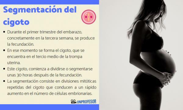
We begin this lesson on the zygote segmentation process by talking about segmentation. The embryonic development It is a complex process that takes a long time, but without a doubt the most interesting part of it is the first third of pregnancy. During the first trimester of pregnancy, specifically in the third week, the
fertilization. At that moment the zygote is formed, which is located in the middle third of the uterine tube.This zygote begins to divide or segment a few 30 hours after fertilization, and this is still covered with a protective layer called zona pellucida. Segmentation is the process of mitotic divisions by which the zygote increases its number of cells.
Segmentation consists of mitotic divisions repeated from zygote which lead to a rapid increase in the number of embryonic cells, called blastomeres or blastomeres.
The segmentation in humans it takes place during the two weeks following fertilization. After segmentation, the gastrulation, a process by which the three germ leaves are established: ectoderm, mesoderm and endoderm, which will give rise to the different human tissues.
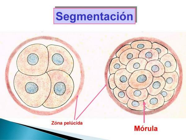
Image: Slideplayer
First week of embryonic development.
Within the zygote segmentation process we will divide the changes that take place in the first and second week of pregnancy. After the two-cell stage, blastomeres divide asynchronouslyIn other words, one of the two blastomeres divides before the other and therefore one of the two masses produced has more cells than the other. As the zygote divides, the cells that form it become smaller with each mitotic division, since there is no increase in mass during this stage of development.
Starting at the nine-cell stage, blastomeres alter their shape and closely align to form a compact cell mass. This process is called compaction and allows a greater interaction between the cells that form the zygote. This close communication is a prerequisite for the next stage: the blastulation. During blastulation, the internal blastomeres separate, which form the embryoblast, and that in later stages they will form the cells of the future embryo.
At this point in development, the zygote already has between 12 and 32 cells and it is called morula (due to its resemblance to a blackberry seen under a microscope). The internal cells of the morula constitute the inner cell mass and the cells that surround them make up the outer cell mass, which will form the embryonic attachments necessary for proper development (for example, part of the placenta).
But, How do cells know in which tissues they have to differentiate? What makes a cell form the embryo or placenta? Very easy, the close contact of the cells with each other, and the different signals they receive, make each blastomeric activate specific genes, which leads to some of them giving rise to parts of the embryo and others to extra-embryonic attachments.
At the end of the morula stage, about 4 days after fertilization, a cavity containing water with sodium ions begins to form between the internal blastomeres. This is called blastocele. and the process by which it appears is called cavitation. In this phase, the embryo as a whole is called blastocyst and its volume remains approximately the same as that of the zygote, except that there is a cavity inside it and it is no longer compact. The end of the blastocyst that contains the inner cell mass is called embryonic poleas it will give rise to the embryo, while the opposite end abembryonic pole.
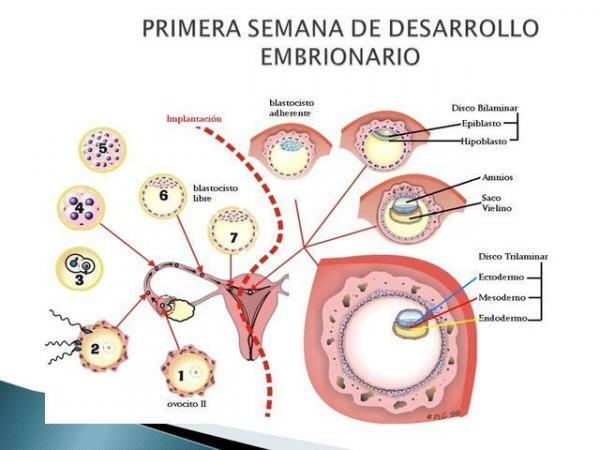
Image: Timetoast
Second week of embryonic development.
The second week of embryonic development is of great importance, since in it the blastocyst that was formed in the last days of the first week will undergo a series of changes that give rise to to the bilaminar embryonic disc (with two sheets or layers of cells), precursor of the three embryonic leaves: ectoderm, mesoderm and endoderm. In addition, while this embryonic disk is forming, the implantation of the zygote in the uterus. In addition, they are formed extraembryonic structures important such as the yolk sac, the embryonic part of the placenta, etc.
The implantation It begins when the blastocyst loses the zona pellucida that covered it and attaches itself to the epithelium of the maternal uterus. At this time, one of the layers of the embryoblast, the trophoblast, begins to proliferate rapidly and gradually transforms into two laminae: an internal one (cytotrophoblast) and a multinucleated outer (syncytiotrophoblast). The syncytiotrophoblast produces enzymes that erode the outermost layer of the uterus, allowing the blastocyst to enter the endometrium.
Once inside the endometrium, the inner cell mass of the blastocyst differentiates into two layers: a layer of small cubic cells adjacent to the blastocyst cavity (hypoblast) and a layer of long cylindrical cells (epiblast). These two layers form a flat disk, a stage of development known as bilaminar embryo.
At this time, within the epiblast we can find a small cavity, which when enlarged constitutes the amniotic cavity. On the other hand, the epiblast cells that migrate and are located adjacent to the cytotrophoblast (amnioblasts) constitute the amnion. Meanwhile, the cells of the hypoblast internally line the blastocele, originating the primitive yolk sac.
Later in development, a new group of cells arises from the yolk sac that forms the extraembryonic mesoderm, this is a connective tissue that surrounds the amnion and the yolk sac. This fabric increases in size and small spaces appear inside; these spaces merge and form a large cavity: the chorionic cavity. This fluid-filled cavity surrounds the amnion and the yolk sac, except in the area where the embryonic disc is attached to the trophoblast through a connecting area called fixation pedicle. Thus, the extraembryonic mesoderm is divided into two parts: somatic extraembryonic mesoderm (covering the amnion and cytotrophoblast) and splanchnic extra-embryonic mesoderm or visceral (lining the yolk sac).
At the same time, the primitive yolk sac begins to narrow, until it is divided into two portions: the largest portion, which is still related to the hypoblast, receives the name of secondary yolk sac, while the other, smaller, remains as a waste that will disappear a few days later.
Now you know how the zygote segmentation process is. If you have any questions about it, leave us a comment and we will answer you!

Image: Slideshare
If you want to read more articles similar to Zygote segmentation process, we recommend that you enter our category of biology.
Bibliography
- Moore, K. L., Persaud, T. V. N., & Torchia, M. G. (Eds.). (2020). Clinical embryology. Elsevier.
- University of Murcia. Master in Biology and Technology of Mammalian Reproduction. (s.f) Course 0: Comparative anatomy and embryology of the reproductive system of domestic mammals. Recovered from: https://www.um.es/documents/9568078/9884658/desarrollo-embrionario.pdf/5b40e5d8-66b1-46ef-9239-2dedafca17a6
- National University of Santiago del Estero (2016) General Embryology. Recovered from: https://www.unse.edu.ar/archivos/ANEXO%20DE%20BIOLOGIA%20Embriologa%20General.pdf