Discover all the STAGES of the ZYGOTE
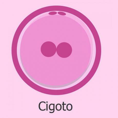
Image: Instagram
The first cell to be produced after fertilization of an egg for a sperm is the zygote. In a previous lesson from a TEACHER we already studied How the zygote forms, but we did not study what happened to this cell when it was formed. In this lesson we will briefly review the stages of zygote and what processes it follows once it has been formed. If you want to know more about the zygote, read on!
Index
- Difference between zygote, embryo and fetus
- Zygote development: segmentation
- Blastocyst formation: blastulation
Difference between zygote, embryo and fetus.
As we already saw in the lesson on the formation of the zygote, the first obstacle that we encounter when studying early development are questions such as when exactly does a zygote cease to be and is called embryo? When should we call the new individual an embryo and when a fetus?
In the human species, these stages are not clearly defined and depending on the author or authors, the distinctions may change slightly. In general, the zygote is that cell, diploid, that is generated as a result of fertilization and undergoes processes of
division, segmentation and blastulation, until producing a structure called blastula or blastocystball-like, a layer of cells that surround a cavity called a blastocele. If you want to know more about the processes and stages that the zygote goes through, keep reading below! In humans, this stage ends approximately one week after fertilization, with the hatching of the blastocyst and the beginning of its implantation in the maternal uterus.On the other hand, the embryo is that initial stage of development from the second week, approximately, to the eighth week. Although its onset is not clear, it is commonly accepted that the completion of the embryonic stage and the beginning of the fetal stage occurs in the passage from the eighth to the ninth week of pregnancy. During this stage, the blastula undergoes the gastrulation process whereby the mass of cells is structured in three layers (endoderm, mesoderm and ectoderm), called gastrula and then organogenesis begins, that is, the formation of the embryo's organs, beginning with the brain, heart and spinal cord and ending with the total formation of the fingers and eyelids.
Finally we find the fetal or fetus stage, no way from the eighth week of pregnancy to delivery. During this stage, the fundamental organs that have begun to form during the previous weeks finish developing and other more structures appear. late but equally important, the fetus begins to store fat and develop certain reflexes such as the grasp reflex (closing of the fingers) or the shock.
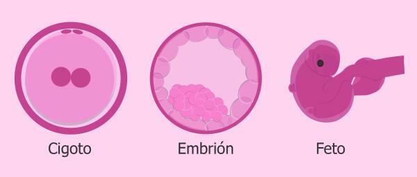
Image: Babygest
Development of the zygote: segmentation.
Once the zygote has formed, it begins to divide, entering the segmentation stage. The segmentation is a process by which the zygote, the cell formed by fertilization, begins to undergo divisions through mitosis successive, one after the other. In this way, the first cell divides into two, each of them divides into two others, forming a mass of four cells and each of those four cells divides into two, etc. This successive division of the zygote gives rise to a mass of cells called morula, which in effect looks like a blackberry. Each of those cells that is produced by mitotic divisions and forms the morula is called blastomere.
It must be taken into account that, depending on the distribution and the amount of yolk contained in the zygote, the divisions during segmentation can be symmetrical or asymmetric and total or partial. In the case of humans we have to remember that yolk comes exclusively from the mother and the divisions occur completely and symmetrically, giving rise to blastomeres that are all the same each.
The morula divides as it progresses through the fallopian tubes, still surrounded by the zona pellucida. It is approximately the fifth day after fertilization, when the morula has about eight blastomeres, when it reaches the neck of the uterus and it continues dividing until it gets about 64 cells.
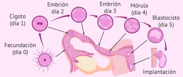
Image: Assisted Reproduction ORG
The formation of the blastocyst: blastulation.
Approximately on the fifth day after fertilization, we have a morula made up of about 64 cells that is already in the cervix. It is now when the hormonal changes occur that allow the blastulation, the cells swell and the zona pellucida finally disappears.
First, the outermost blastomeres will become thinner and bond tightly together, forming a layer of peripheral cells of the blastocyst that from now on we will call trophoblast. The cells of the trophoblast will in the future form the embryonic placenta and the umbilical cord, which allow nutrition and gas exchange between the fetus and the mother.
At the same time as the trophoblast, cavitation occurs. The cavitation It is a process by which an accumulation of cells occurs in the blastomeres in the center of the morula. water and ions and makes a cavity appear, filled with liquid like a water-filled balloon, call blastocele.
Also simultaneously, between the trophoblast and the blastocele and at one of the poles, a mass of cells called embryoblast. This mass of blastomeres will be the one that, in the future, will give rise to the embryo. In this mass of cells we can differentiate two layers that will be very important:
- Hypoblast: is the layer of the embryoblast that is between the embryoblast and the blastocele (the cavity of the blastula). This layer of cells will form the part of the primary yolk sac.
- Epiblast: it is the layer of the embryoblast just below the previous one, which are in contact with the hypoblast on one side and the trophoblast on the other. This part of the embryoblast is adjacent to the amniotic cavity.
Once the blastocyst is formed, approximately six or seven days after fertilization, it approaches the outer layer of the uterus, called endometrium and adheres to it, usually in the area closest to the embryoblast. As soon as it contacts the endometrium, the trophoblast cells begin to divide and begin to move into the uterus. This process is called implantation. The entry of the blastocyst into the uterus does not end until a week later, 14 days after fertilization. It is from the third week, when the blastocyst completes its implantation, when some authors locate the passage from zygote to embryo.
And with this we end this lesson with the stages of the zygote so that, thus, you better understand what human fertilization is like.
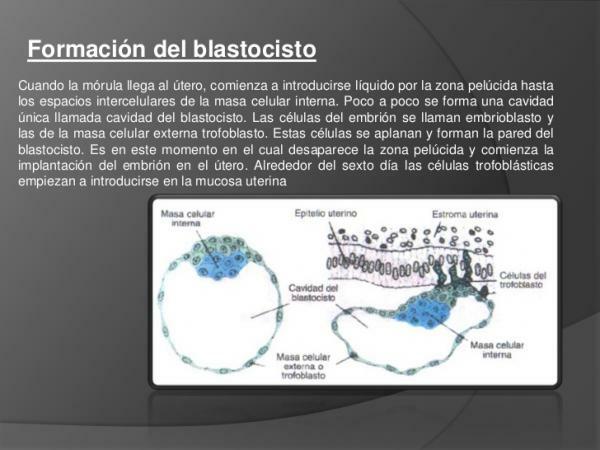
Image: Slideshare
If you want to read more articles similar to Stages of the zygote, we recommend that you enter our category of biology.