INTERNAL ear: parts and functions

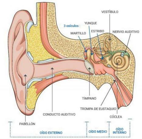
The inner ear is responsible for transform sound waves, which the middle ear has transformed into vibrations, in electrical impulses that the brain interprets as sound sensations. In addition it is also the organ of balance, since, together with the sight and the musculoskeletal system, it is responsible for the sense of balance (equilibrium-perception). The equilibriumception, is a physiological sense of great importance that gives us the ability to register imbalances and correcting them in order to maintain body stability both in movement and in position static. In this lesson from a TEACHER we explain in detail the parts and functions of the inner ear.
Index
- The bony labyrinth, one of the parts of the inner ear
- The membranous labyrinth, another part of the inner ear
- Functions of the inner ear
The bony labyrinth, one of the parts of the inner ear.
Unlike outer and middle ear, the inner ear is immersed in a liquid medium
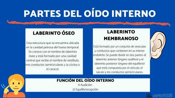
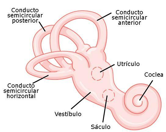
. Two structures are distinguished at the level of the inner ear: a bony part and a part formed by membranous structures. The bone structure is the bone maze which is filled with fluid and contains the membranous maze, which are the membranous organs; that reproduce the shape of the bone structure that contains them and that float in their internal fluid.The bony labyrinth is one of the parts of the inner ear. This structure that is located in the petrous cavity of temporal bone (bone of the medial lateral zone of the base of the skull). This cavity is known by the name of bone maze and is formed by: a central cavity that receives the name of vestibule, three semicircular canals, and the cochlea or the snail.
A series of membranous organs are housed in the bone labyrinth that follow the contour of the bone labyrinth and which, as a whole, is known as membranous maze. The bony capsule of the labyrinth is filled with a fluid, the perilymph, which protects the membranous structures that form the membranous labyrinth. These membranous structures float in the perilymph. The perilymph has a composition similar to the extracellular medium.

The membranous labyrinth, another part of the inner ear.
We continue to know the parts of the inner ear to speak, now, of the membranous labyrinth that is formed by a set of vesicles and ducts that contain inside endolymph. The membranous labyrinth can be divided into two parts: the anterior labyrinth (auditory organ) and the labyrinth posterior (organ of balance) that is composed of the utricle, the saccule and the semicircular canals.
They are the structures in charge of transform mechanical stimuli transmitted through the middle ear in electrical impulses and initiate the sending of nerve signals to the cerebral cortex. The cochlea transmits information about sounds and the utricle, saccule, and semicircular canals inform about balance.
Anterior membranous labyrinth: The organ of hearing
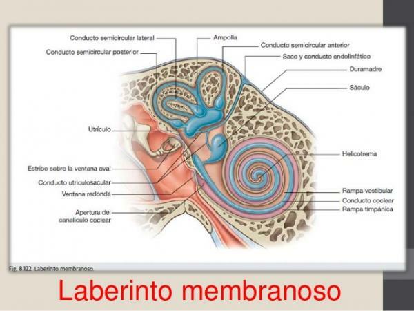
The membranous duct, or anterior labyrinth, is a coiled tube, of about 35mm approximately, that is inside the cochlea or the bony snail and that receives the name of cochlear duct. The spiral tube is wound around a bone axis called modiolo. The cochlea communicates with the middle ear through two orifices closed by membranes: the oval window and the round window.
The cochlear duct is divided into three cavities: the vestibular ramp, the cochlear duct or middle ramp and the scala tympani.
- Vestibular ramp or scale: It is full of perilymph, and connects with the oval window membrane (at the base of the buccal ramp), through which the stapes transmits its vibration to the oval membrane. This cavity connects with the tympanic cavity in the helicotrema (at the apex of the spiral of the membranous cochlea).
- Colear duct, ramp or medium scale: This cavity is located in the intermediate zone, it is filled with endolymph and is separated from the upper cavity (buccal ramp) by a membrane that is called the vestibular window or Reissner's membrane.
- Tympanic ramp: The separation with the other cavity, the scala tympani, is the basilar membrane. This membrane has a tonotopic organizationIn other words, it is a membrane made up of neurons that respond to specific sound frequencies. On the surface of the basilar membrane is located the organ of Corti. It is the neurosensory organ of the cochlea, in which the transformation of the mechanical impulse transmitted by the waves generated by the vibration of the oval window membrane caused by the stirrup, in pulses electrical
The organ of Corti is composed of sensory or ciliated cells, nerve fibers connected to them, and supporting structures. The hair cells present in their apical area (upper end) stereocilia. These cells are connected to neurons whose axons constitute the auditory nerve. The organ of Corti contains about 20,000 hair cells.
Ramp or tympanic scale: It is the third compartment of the membranous cochlea and borders the basilar membrane. At the base of this cavity is the round window that communicates with the middle ear through a thin membrane. This cavity is filled with perilymph, as in the case of the buccal ramp.
The posterior labyrinth
- Otolithic organs (Utricle and Sacculum): These two membranous structures of the inner ear are responsible for informing the brain of which is the head position at all times. Is about two cavities located between the semicircular canals and the cochlea. They are full of endolymph that communicate with the semicircular ducts (utricle) and with the cochlea (saccule) .These two bags have walls lined with stereocilia that are covered by a gelatinous matter that contains calcium carbonate crystals: the otoliths. The force of gravity causes the movement of the otoliths when the head moves, and these cause the movement of the stereocilia that stimulate the neurons connected to them. Neurons, whose axons are part of the auditory nerve, send nerve signals to the brain to indicate at all times what is the position of the head.
- Semicircular canals: These structures inform the brain of the direction of movements of head turn. It is about three semicircular canals with different orientation in space, more or less at right angles to each other. The inside of the ducts is filled with endolymph. The five orifices of the semicircular canals communicate with the vestibule. In each of the ducts there is a gelatinous structure called Dome, which covers a group of hair cells. The movement of the dome in response to the movements of the head, causes the movement of the stereocilia of the hair cells that send signals, through the auditory nerve, to the brain indicating the orientation of the turn of the head. The brain integrates the information provided by each of the semicircular canals that have different spatial orientation.

Functions of the inner ear.
As we have already seen in the sections that explain what the parts of the inner ear are, it fulfills two fundamental functions:
Hearing
It is one of the main functions of the inner ear. The part of the inner ear responsible for hearing is the anterior membranous labyrinth. When the middle ear, through the stapes, transmits the vibration caused by sound, the membrane of the oval window causes the formation of waves in the endolymph that fills the cochlear duct.
The endolymph waves in turn cause the movement of the stereocilia of the hair cells of the organ of Corti, which are connected to a group of neurons that convert the mechanical movement of stereocilia into nerve impulses. The axons of these neurons connected to the hair cells, unite to form the nerve auditory, responsible for transmitting nerve signals to the cerebral cortex where they are interpreted as sounds.
Hearing is a subjective experience, it is the mental representation of the immediate sound environment. The hair cells of the organ of Corti, together with the neurons connected to them, are capable of inform the brain of the frequency, intensity and orientation of the perceived sounds.
- The frequency of sound allows us to distinguish between bass and treble sounds. As we have already commented previously, the organ of Corti contains a tonotopic map composed of neurons that react specifically at a certain frequency. The periodicity with which nerve impulses are generated in the hair cells also helps to determine the tone of the sound. Treble tones have a higher periodicity than bass tones.
- The intensity and it allows us to distinguish if a sound is strong or weak. This sensation will be defined by the intensity of the pressure that the sound wave exerts on the eardrum. A threshold of hearing is established, the minimum pressure so that a sound can be perceived and a threshold of pain that is the pressure from which the hearing produces a sensation of pain.
- The orientation of sound is possible thanks to binaural hearing, that is to say, each of the ears perceives the sound, and allows the localization of the sound in the horizontal plane. It is the difference in intensity and time in the perception of sound by the two ears that allows locating the location of its origin.
Equilibrioception
It is another function of the inner ear. The balance organ of the inner ear is the vestibular system or posterior labyrinth. In it the hair cells of the semicircular canals report the movements of the head. Whereas, the stimulation of the hair cells by the movement of the otoliths indicates the position of the head as a function of the force of gravity.
When the body is in motion, the vestibular system detects gravity and other mechanical forces that stimulate the hair cells of the semicircular canals and the otolithic organs. This body acts by coordinating with other systems that contribute to balance such as vision and neuromuscular proprioception system, to control the position of the body at rest or at movement. This helps maintain a stable posture and maintain balance during the execution of movements such as walking or running. It also helps keep visual focus stable from surrounding objects when the body changes position.
Failure in the vestibular system causes feelings of instability, dizziness and vertigo attacks.

Image: Cotral Lab
If you want to read more articles similar to Inner ear: parts and functions, we recommend that you enter our category of biology.
Bibliography
Elaine N. Marieb (2008). Human anatomy and physiology. Madrid: PEARSON EDUCACIÓN S.A.


