CENTRIOLOS: functions, characteristics and structure

Throughout an individual's life, their cells are dying and in order to maintain themselves, they need to divide into two daughter cells during the process of cell division. However, to carry out this process properly it is necessary that a series of cell structures fulfill their function correctly. Among these structures is the mitotic spindle, which arises from structures known as centrioles. In this lesson from a TEACHER we will see what are centrioles, their functions, characteristics and structure. Read us below to learn more about them!
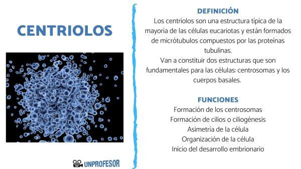
Centrioles are a typical structure of most of the eukaryotic cells and they are made up of microtubules composed of tubulin proteins.
The centrioles will in turn constitute two structures that are fundamental for cells, such as Centrosomes that act in cell division and basal bodies that form the cilia and flagella, structures that perform different functions.
Both the centrioles and the basal bodies have the same molecular structure and they are interchangeable
in the cell, that is, the centrioles can move to the membrane to form cilia and the basal bodies can move into the cells and form a centrosome.The function of the centrioles in the centrosome is that of organize them, while its function in the basal bodies is to organize and begin to form the microtubules that will form the axoneme or skeleton of cilia and flagella.
In human eukaryotes, the mature centrioles or basal bodies are cyclindrical structures with between 150 and 500 nm in height (it is more variable and it is not known how it is established) and about 250 nm in diameter, for so much, the centrioles and basal bodies are two of the largest protein structures of the eukaryotic cell.
The walls of the centrioles are formed by nine microtubule triplets arranged longitudinally and oriented all in the same direction, with the ends over the microtubules forming a part of the cylinder and the ends less in the other, forming a distal and a proximal end of the centriole or basal body, that is, they are structures polarized. However, this structure not fulfilled in all organisms, as for example in some fly embryos, where there are 9 pairs, or in the nematode C. Elegans, where there are 9 simple microtubules.
In the microtubule triplet, only one is complete and is made up of the 13 protofilaments (formed by 13 tubulin filaments assembled together). This complete microtubule is called microtubule A, while microtubule B and C are incomplete and only consist of 10 protofilaments, sharing 3 with those of A. At the distal end of the centriole, only microtubules A and B reach and C is shorter. At the proximal end in the young centrioles a cart-like structure is formed and helps to organize and assemble the 9 microtubule triplets.
The centrosomes of the cells are structures formed by two centrioles, one mature and one immature. The mature centriole has protein structures that will constitute distal and sub-crystal appendages and it is the distal appendages that are associated with the plasma membrane. to form the basal bodies of the cilia at the time that the mature centriole migrates to the vicinity of the membrane, while the sub-crystals are responsible for anchoring the microtubules.
The basal bodies too have a kind of appendix at their distal ends, but in this case they are called basal feet and connecting or transition fibers, while at their proximal end they have striated ciliary roots. These appendages help the basal body to anchor itself to the plasma membrane, and the striated roots help organize the cellular structure of the basal body.
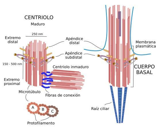
Image: Atlas of Plant and Animal History
The centrioles have several functions for the eukaryotic cell and for its proper functioning. Among these functions are the following.
Formation of the centrosomes
Centrosomes are the main elements of animal cells that serve to begin to form microtubules of the cytosol, process known as microtubule nucleation. A centrosome is made up of a pair of centrioles (one mature and one immature) surrounded by a cloud of molecules that form the pericentriolar material. The evidence shows us that the centrioles may be responsible for assembling the centriole, since they are the ones that recruit the pericentriolar material and the rings of the gamma subunits of the tubulin protein that are in the pericentriolar matrix and seem to be those that really serve for the nucleation of the microtubules
The centrioles and the pericentriolar material that surround them play one of the most important roles during cell division of animal cells, since they are responsible for constitute the mitotic spindle. However, it is not the same in all cells and it has been seen that in neurons, epithelial cells, and muscle cells, the centrosome is not the main microtubule nucleator. Centrosomes are also absent in plant and yeast cells, where the mitotic spindle it is constituted in the absence of centrioles.
Cilia formation or ciliogenesis
The ciliaare Mobile or non-mobile processes of the surface of the plasma membrane of some eukaryotic cells. They perform important functions where they are, such as defense against microorganisms and mucus movement on the respiratory surfaces, displacement of the oocyte formed by the fallopian tubes or sensory functions in the auditory apparatus and other sense organs. Its formation occurs from the basal bodies by elongation that occurs by polymerization of microtubules A and B of each of the triplets.
When a cell finishes its cell division, an old centriole migrates towards the plasma membrane and becomes the cilia-forming basal body. There are cells that have thousands of cilia on their free surface, such as the cells of the trachea, the ependyma or the oviducts. For the formation of independent cilia in these cells, the basal bodies must migrate to the cell surface and other elements of the cytoskeleton such as actin microfilaments and microtubules.
Since the presence of cilia is not compatible with the division of the cell, these must be disassembled when the cell is going to divide and assembled again once this process is finished. This disassembly is believed to occur so that the basal bodies do not interfere with the centrioles during mitotic spindle formation.
Cell asymmetry
In asymmetric divisions there is an unequal distribution between both daughter cells and the centrioles are necessary for this type of division, since they will contribute to the correct orientation of the mitotic spindle. Another way to create an asymmetry depends on which daughter cell takes the oldest centriole.
The oldest centriole seems to surround itself with molecules slightly different from those that surround the youngest, serving the stem cells to distribute among the daughter cells the different molecules associated with the pericentriolar material of one or the other of the centrioles, such as messenger RNA or the different factors that help start and maintain transcription.
One hypothesis that has been observed is that the cell that manages to capture the centrosome that has the oldest centriole ends up first developing the cilia that They serve to respond earlier to the different signals in the environment, that is, this uneven distribution can produce a different behavior between the two cells daughters.
Cell organization
The position in which the centrioles are located in the cytosol of the cells, constituting the centrosomes of the cells, are important for determine the organization of many cells, or to allow cell movement, since they help to create a differentiation between the advancing front and the back of the cell. For example, in central nervous system astrocytes (cells that help neurons), the Golgi apparatus it is arranged towards the advancing front of the cell due to the action of the centrosome. However, in fibroblasts the cell nucleus is arranged in the most caudal part of the cell, also thanks to the action of the centrosome.
The position of both the centrioles and the centrosome in cells appears to be determined by the interaction between microtubules and actin microfilaments. It has been seen that the position of the centrosome in the cell depends on the interaction between the microtubules it produces and the cell cortex that is located on the inner face of the plasma membrane and is made up of microfilaments of actin.
However, sometimes the centrosome is located in the vicinity of the cell nucleus due to the interaction with proteins that are part of the nuclear envelope and anchor it in this position. In other eukaryotic cells, the relationship between centrosomes and nuclear envelope are due to fibers proteins associated with the centrioles called striated fibers, which serve as a union for both structures.
Beginning of embryonic development
After the fusion of the two haploid cells in the fertilization process, only the spermatozoonwill remain with a centriole which comes from the basal body of the flagellum. This centriole will recruit the pericentriolar material found in the ovule to form the centrosome.
This newly formed centrosome will take care of the nucleation and organization of the microtubule system cells necessary for the migration and fusion of the two pronuclei (haploid nuclei of both gametes). Later, it will divide and form the mitotic spindle responsible for carrying out the first division of the cell.
If you want to read more articles similar to Centrioles: functions, characteristics and structure, we recommend that you enter our category of biology.