
Neuron functions
The neurons they are one of the most interesting and fascinating cells in our body. One of the most defining characteristics of neurons is their structure and their parts. The parts of a neuron are three: body, dendrites and axon. The appearance of these three parts within neurons is not a whim of evolution, but has to do with the function that each one of them carries out within the neuron. In this lesson from a TEACHER we will explain more about the neuron functions within the transmission of the nerve impulse.
Index
- Main functions of the neuron
- The soma or neuronal body
- Neural dendrites
- The axon, another part of the neuron
- Other structures and cells associated with neurons
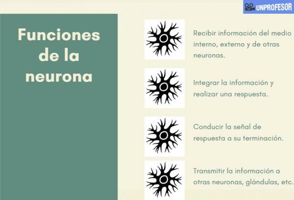
Main functions of the neuron.
Neurons are one of the main cells of the central nervous system: brain and spinal cord. The functions of the neuron is that of within it is that of receive, process and conduct information through chemical and electrical signals.
This is carried out thanks to the
electrical excitability of your plasma membrane, one of the most special and defining characteristics of neurons. A typical neuron consists of:- Body or soma. Within it there is a central nucleus, very voluminous and normally very active, and the perikaryon or cytoplasm, which surrounds the nucleus and where the cellular organelles are found.
- Neurites: Usually an axon and several dendrites emerging from the body or soma.
The soma or neuronal body.
To better understand the functions of the neuron, it is important to discover the different parts by which it is formed. The soma or neuronal body produces the energy and substances necessary for the functioning of the neuron. Within the neuronal soma we can find two well-defined parts:
- Core.
- Pericarion.
In the center of the neuronal soma a core central with one or two prominent nucleoli and a dispersed, transcriptionally very active chromatin. Within this the interpretation of the information received by the neuron and the generation of the exit command. This starting command will normally be the release of a neurotransmitter or an electrical impulse.
Surrounding the nucleus is the perikaryon. The perikaryon It is the specific name given to the cytoplasm of the neuron. The perikaryon is rich in organelles and is very active. Among the most important organelles, the corpuscles of Nissl stand out.
The bodies, corpuscles or substance of Nissl they are free ribosomes adhered to the rough reticulum. Its function is the synthesis of proteins, hence its great importance and number. Under certain conditions, both normal and pathological, there may be fewer Nissl bodies, indicating decreased production of neuronal proteins.
Within the perikaryon we also find the Golgi apparatus, lysosomes, mitochondria, etc.. in charge of other no less important functions such as energy production, waste elimination, maturation of proteins generated by Nissl bodies, etc.
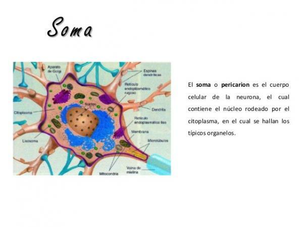
Image: Slideshare
Neural dendrites.
Dendrites are another part of the neuron. Is about cytoplasmic projections of the neuron that are surrounded by a plasma membrane but without myelin envelope.
Its function is the transmission of information to the neuron, that is, neurons are the information entry way from a structure, organ, etc. to the interior of the neuron.
Like soma, dendrites have structures features:
- They have many microtubules and few neurofilaments. Both microtubules and neurofilaments are arranged in parallel.
- They have many mitochondria. They need a lot of energy, so in the dendrites we find many mitochondria.
- Nissl corpuscles. Nissl corpuscles are more abundant in the area adjacent to the soma, although they can also be found in other parts of the dendrite.
- Smooth endoplasmic reticulum. Abundant smooth endoplasma reticulum, especially in the form of synapse-related vesicles, areas of information exchange with other neurons (intermediate neurons)
In the terminal portion of the dendrites, the furthest from the soma, we can find extensions called spines. The dendritic spines they are small, actin-rich membranous structures that protrude from the dendrite and facilitate contact of the dendrites.
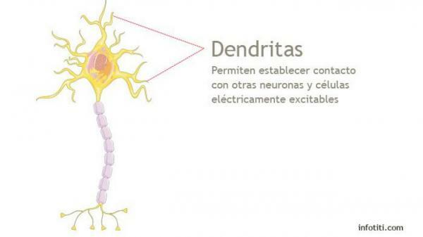
Image: Titi
The axon, another part of the neuron.
We continue to analyze the functions of the neuron, now talking about the axon that is a prolongation of the neuronal soma, longer or more extensive than the dendrites, which is surrounded by its membrane (axolema). The axon can be divided into three parts: the axon cone, the initial segment, and the rest of the axon.
The axolema can be covered or devoid of the myelin sheath, a structure that, as we will see later it helps the transmission of the nerve impulse and that appears in organisms more developed.
In the terminal portion of the axons, further from the soma, we can find the synaptic buttons. Synaptic button is the name given to the final part of the axon, which divides to produce different endings that produce synapse with other neurons.

Image: Slideshare
Other structures and cells associated with neurons.
To perform its function, neurons are not alone. As we will see below, it is important that neurons have other cells that help them:
- The myelin sheath. The myelin sheath is a thick layer of protein and fatty substances that accumulates around the axons, similar to the protective plastic layer of electrical cables. This covering allows nerve impulses to reach longer distances, without losing intensity.
- Ranvier's nodules. Ranvier's nodules are periodic thinning or narrowing of the myelin sheath that surrounds the axon. Its function is that the nerve impulses travel with greater speed and appear in the final part of the axon.
- Schwann cell or neurolemocytes. Schwann cells are responsible for synthesizing and repairing the myelin sheaths.
- Glial cells. They are cells that provide structural and metabolic support to neurons. Some of the most important are astrocytes (star-shaped), ependymal cells, microglia (immune) cells, or oligodendrocytes.
If you want to read more articles similar to Neuron functions, we recommend that you enter our category of biology.
Bibliography
- Paniagua, R., Nistal, M., Sesma, P., Álvarez, U. M., Fraile, B., Anadón, R.,... & De Miguel, M. P. (1998). Plant and animal cytology and histology (No. QH573. C57 2007.). McGraw-Hill Interamericana.
- Bologna, C (June 5, 2017) What are the parts of a neuron? Recovered from https://www.lareserva.com/Cuales_son_las_partes_de_una_neurona


