Structure of the NEURON -SUMMARY + SCHEMES !!
Neurons are one of the more complex cells and special of the human body. For years, neurons have fascinated man and have been the object of study at different levels.
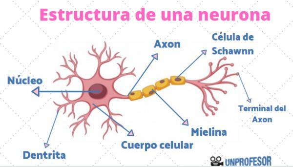
In this lesson from a TEACHER we will address neuron structure, that is, we will see the different parts that we can differentiate in a neuron: body or soma, nucleus, axon, dendrites, myelin sheath, substance of Nissl, nodules of Ranvier, synaptic buttons and cone axon. If you want to know more, we invite you to continue reading!
Index
- What is a neuron?
- The body or neuronal soma
- The nucleus of the neuron
- The neuronal axon
- Dendrites
- The substance or bodies of Nissl
- Myelin sheaths
- Ranvier's nodules
- Synaptic buttons
- The axon cone
What is a neuron?
The neurons are the structural and functional unit of our brain, that is, they are the minimum structurally differentiable part that retains its own function.
Neurons are also called brain nerve cells, since They are the cells that are the main cells found in the nervous system, although they are not the only ones. The
neuron functionIt is to process, store and transmit the information that we receive from the outside, and that is why they fascinate us so much. Neurons are able to communicate through a process of chemical and electrical signals that can be connected through neurotransmitters, that is, the messenger that is responsible for transmitting the information between each neuron.In order to perform its function, the neuron has different areas within itself, which are responsible for performing different functions. But be careful, as in a chain manufacturing factory, the function of each of the parts will depend on the others doing their function correctly.
The body or neuronal soma.
We are already beginning to know the structure of the neuron to refer to the neuronal body or neuronal soma, that is the center or 'body' of the neuron; If you observe it through a microscope, this part can be differentiated well, since it is the widest part of the cell and has a characteristic flower or star shape.
The neuronal body is the place where the neuron metabolic activity, that is, where all the electrical processes that allow the transmission of information occur. Also, it is the place where the genetic material is formed for its cellular survival (cytoplasm), through the generation of proteins and contains different types of cellular organelles that allow its maintenance.
The nucleus of the neuron.
Like any cell, another of the parts of neurons is he core. The nucleus of the neuron is located inside the soma and is a structure delimited from the rest of the cytoplasm by a membrane. Inside you will find protected DNA, that is, the genes of the neuron.
The nucleus is in charge of controlling the expression of genetic material and, therefore, regulating everything that happens in the neuron. The nucleus of the neuron is, as in the rest of the cells, the command center of the cell.
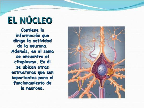
Image: Slideshare
The neuronal axon.
The axon is a single extension that is born from the body or soma of the neuron, that is to say, that for each neuron we will only be able to find one axon. The axon is located opposite the dendrites and is responsible for drive electrical impulse even the synaptic buttons once the neurotransmitters have been received and the body has been electrically activated. Therefore, the main function of the axon is to release neurotransmitters, which will inform the next neuron of what to do.
Therefore, the axon is a unique tube that arises from the body of the neuron and that, unlike dendrites, does not capture information, but is already directed to transmit it.
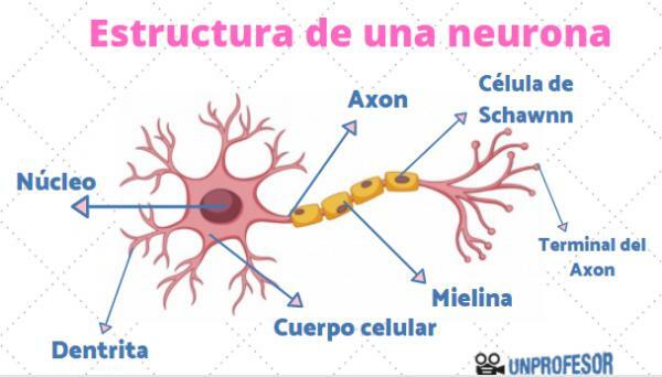
The dendrites.
The dendrites is another of the parts that make up the structure of the neuron. They are extensions of the neurons that are born from the body or soma and that make up a kind of branches that cover the entire center of the neuron. That is why sometimes the set of dendrites of a neuron is called a dendritic tree.
The main function of dendrites is to capture neurotransmitters produced by the nearest neuron and send the chemical information to the body of the neuron to make it become electrically activated.
Therefore, the dendrites are the ramifications of the neuron that they capture the information in the form of chemical signals and transmit to the body the information received from the previous neuron in the network. This neuron can transmit information from the sensory organs or the viscera to the brain or vice versa, from the brain to the organs.
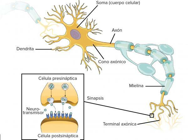
Image: Khan Academy
The substance or bodies of Nissl.
The Nissl substance or Nissl bodies are a set of granules found in the cytoplasm of neurons, both in the body and the dendrites, but not in the axon.
Nissl's bodies take care of make proteins, mainly responsible for the correct transmission of electrical impulses.
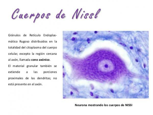
Image: Slideshare
Myelin sheaths.
Although less known than the previous ones, the myelin sheaths are very important structures for neural function, since they take care of facilitate the passage of information generated in the soma. The myelin sheaths allow the electrical impulse to flow without any problem within the axon, since they are a kind of capsules made of protein and fat, which cover the axon until reaching before the synaptic buttons.
The myelin sheath of neurons is not continuous throughout the axon. In fact, myelin forms "packs" that are slightly separated from each other.
When there is a problem in the production of myelin, the transmission of information slows down, that is, electrical impulses and responses are slowed neurons, as they cannot travel their path with the proper speed. These problems are those that appear in neurodegenerative diseases typical of advanced age.
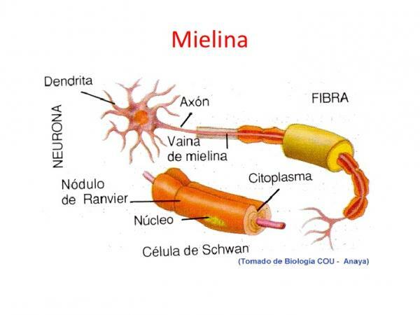
Image: Slideshare
Ranvier's nodules.
As we have seen previously, the myelin sheath was not continuous. This gap, which is less than a micrometer in length, is what is called Ranvier's nodule.
Ranvier's nodules are small regions of the axon that are not surrounded by myelin and that expose it to the extracellular space. These free spaces are essential for the transmission of the electrical impulse to occur normally, since at sodium and potassium ions enter through it, essential for the electrical signal to travel faster through the axon.
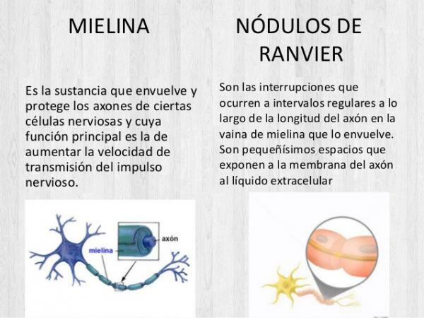
Image: Slideshare
Synaptic buttons.
Synaptic buttons are located at the end of the axon. At the end of the axon we can see that it is divided into two fragments: these small branches have small bumps, quite similar to dendrites, which we call synaptic buttons.
These synaptic buttons are in charge of release neurotransmitters with the responses generated in the soma and produced by the axon, so that the nearest neuron receives it.
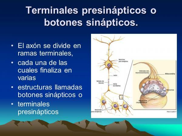
Image: Slideplayer
The axonal cone.
If we observe the neuron under a microscope, we will not be able to differentiate the axonal cone with the naked eye, that is, it is not a structurally differentiated part of the neuron. For this reason, its existence is often not discussed in reviews of the structure of the neuron, despite being a key part of its functioning.
The axon cone is the region of the body of the neuron that narrows to give rise to the axon. This area is very rich in channels and conveyors They require energy (in the form of ATP), so this area of the neuron has a high concentration in mitochondria. In addition, it is also very rich in ions, since these have to be transported from one area of the neuron to the other.
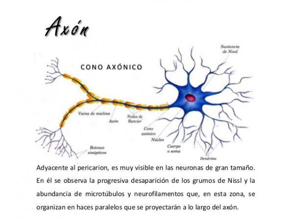
Image: Slideshare
If you want to read more articles similar to Neuron structure, we recommend that you enter our category of biology.
Bibliography
- Bertran Prieto, P (s.f) The 9 parts of a neuron (and their functions). MedicoPlus.
- Bolonia, C (June 5, 2017) What are the parts of a neuron?. The reserve.
- Cuesta, E (s.f). The 9 parts of a neuron (and their characteristics). Next style.

