Anatomy of the human eye: parts and functions
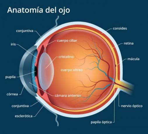
Image: All About Vision
The view is the most important sense of human beings. Almost 50% of the brain is dedicated to visual processing. The main organ involved in vision, after the brain, is the eye. The eye is an organ that, despite appearing very simple at first glance, has a very complex structure made up of eleven parts, each with a very specific function. If you are interested in learning more about this organ, you just have to continue reading this lesson from a PROFESSOR in which we will review the human eye anatomy with all its parts and functions.
Index
- What is the eye?
- The cornea, an important part of the anatomy of the eye
- The iris, another part of the anatomy of the eye
- The pupil of the human eye
- The lens
- Aqueous humor
- Sclera
- The conjunctiva
- The choroid
- The vitreous humor
- The retina
- The optic nerve
What is the eye?
The eye is a structure capable of capturing light and convert information into electrical impulses that travel through neurons to the brain, where the information received is processed. In higher organisms such as humans, the eye is a very complex system capable not only of
catch the surrounding light, but of regulate its intensity through a diaphragm (iris) and focus the objective thanks to an adjustable lens structure (crystalline) to form the image.Later, this image turns it into a electrical sign set that reach the brain through complex neural pathways that connect, via the optic nerve, the eye to the visual cortex and other brain areas important for the processing of this information.
To do all of these things effectively, the eye has many structures, with different parts of the eye and with different functions. If you want to know more about the anatomy of the human eye, read on!
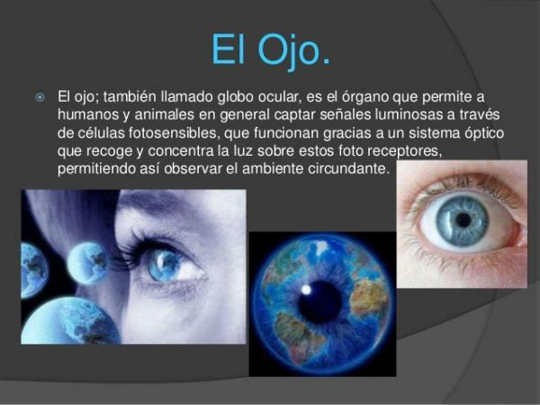
Image: Slideshare
The cornea, an important part of the anatomy of the eye.
The cornea It is part of the anatomy of the human eye. It is the part of the eye that is in contact with the outside. It is domed in shape and approximately one millimeter thick. The cornea is a transparent layer that covers the iris and lens and allows the refraction of light, that is, that light penetrates into the interior of the eye. To function properly and protect it from the outside there are tears and aqueous humor.
When the cornea is altered and does not have the same thickness in all its parts, a vision problem called astigmatism occurs. The astigmatism affects near and far vision, and makes objects appear blurry or distorted. This can be easily corrected with the use of glasses, contact lenses or by means of a more or less simple operation with laser or implantation of a lens inside the eye (intraocular).
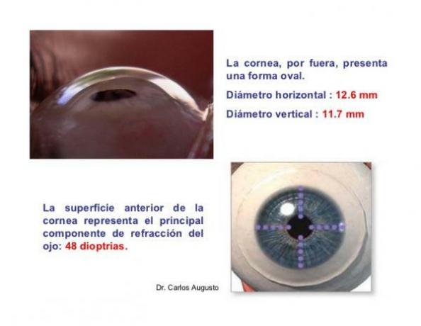
Image: Slideshare
The iris, another part of the anatomy of the eye.
The iris is a circular membrane that separates the anterior and posterior chambers of the eye. This membrane is controlled by two muscles, which regulate the amount of light that enters the eye. On the one hand, the pupil dilator muscle makes the pupil larger (pupil dilation or mydriasis) and more light enters the eye; the sphincter muscle of the pupil causes the pupil to be narrowed (miosis) and less light to enter.
The iris tissue is pigmented thanks to the presence of different types of melanin at different depths of the iris, which cause the different colors of the eyes. Eye color is genetically determined and is reached between six and ten months of age.

Image: Slideshare
The pupil of the human eye.
The pupil It is a hole in the center of the iris that allows light to pass through to the posterior chamber of the eye. It appears as a black circle in the central part of the eye that can have a variable diameter between people who but which is usually between 3 and 4.5 millimeters in normal conditions and can widen up to 9 mm in the darkness.
Its function is, together with the iris, regular through muscle contractions the amount of light that enters the retina.
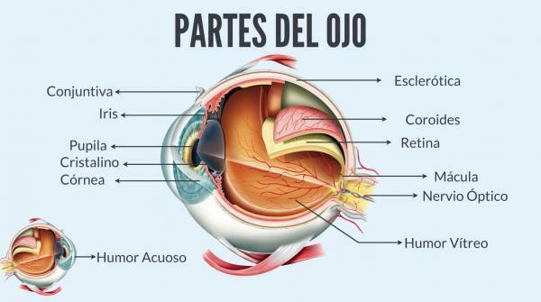
Image: Brill Pharma
The lens.
Within the anatomy of the human eye we also find the crystalline which is the biconvex lens that is located behind the iris and allows vision focus. Accommodation is the process by which the curvature and thickness of the lens are modified to focus on objects based on their distance from the person. When light rays pass through the lens, they are concentrated in a part of the retina and the image is formed.
Related to the lens we can find two alterations: presbyopia and cataracts. As we age, the lens progressively loses its ability to accommodate as it hardens. This phenomenon is known as presbyopia or eyestrain. On the other hand, the loss of transparency of the lens, which can affect vision, is called waterfalls. Cataracts can appear due to the use of certain medications or simply due to aging. Both alterations are corrected with the use of glasses and, in the case of cataracts, with a simple operation.
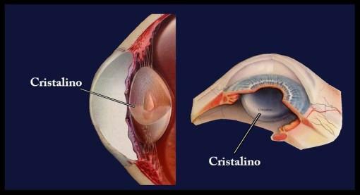
Image: Ophthalmology-online
Aqueous humor.
The aqueous humor It is a clear liquid that is between the cornea and the lens. The cornea and lens do not have blood supply, so it is the aqueous humor that is responsible for nourish to these two structures, oxygenate them and allows the eye pressure stay constant. This liquid is made up of water, glucose, vitamin C, proteins, and lactic acid.
Aqueous humor is being generated and eliminated continuously, every 90 minutes or so. The imbalance between the two can cause pathologies to appear. If the pressure of the aqueous humor rises, a disease known as glaucoma.

Image: Ophthalmology-online
The sclera.
The sclera It is a thick and resistant membrane that covers the eyeball, giving it its characteristic white color and protecting internal structures. The anterior part of the sclera is attached to the cornea, while the posterior part has an opening that allows the connection between the optic nerve and the retina.
The conjunctiva.
The conjunctiva It is a transparent mucous membrane that lines the sclera and the back of the eyelids, which is in contact with the cornea. Contribute to the lubrication and disinfection of the eyeball since it produces tears and mucus, although the lacrimal glands are more relevant in this regard.
Being in contact with the outside, trauma, infections and allergic reactions can occur in the conjunctiva, which can become inflamed and lead to conjunctivitis.
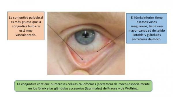
Image: SlideShare
The choroid.
The choroid It is the membrane lined with blood vessels and connective tissue that separates the retina and the sclera. The choroid provides the retina with nutrients and oxygen it needs to function properly, in addition to maintaining a constant temperature in the eye. In addition, it is dark in color, so it prevents light from reflecting or bouncing inside the eye.
The vitreous humor.
The posterior chamber of the eye, between the lens and the retina, is filled with vitreous humor. The vitreous humor It is a transparent, gelatinous liquid with a density greater than that of the aqueous humor in the anterior chamber. It constitutes most of the eyeball and its functions are to provide it with rigidity, cushion impacts, maintain intraocular pressure and fix the retina.
Unlike the aqueous humor, the vitreous humor is generated during the embryonic stage and is not renewed, so it ages as the individual does. Sometimes the liquid darkens due to the rupture of a vein or waste substances appear that cannot be eliminated by phagocytes, causing shadows in the vision (floaters or myodesopsias).
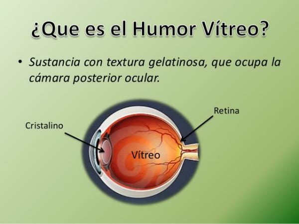
Image: SlideShare
The retina.
The retina It is also part of the anatomy of the human eye and it is a thin and partially transparent layer that is in contact with the inner face of the choroid and with the vitreous humor. The retina is the true receiving organ of the visual system since in this structure the photoreceptors (rods and cones). This membrane lines the back of the eye and has a function similar to that of a screen: the lens projects the images perceived in the retina, the rods and cones capture it and it is transmitted to the brain through the nerve optical. An area of the retina, called the fovea, is responsible for detailed vision as it has a large concentration of cones.
There are numerous diseases related to the retina, but the main ones are retinal detachment (separation of the layer of neurons in the epithelial layer (skin) to which they are normally attached) and retinitis pigmentosa (progressive loss of cones and Canes).
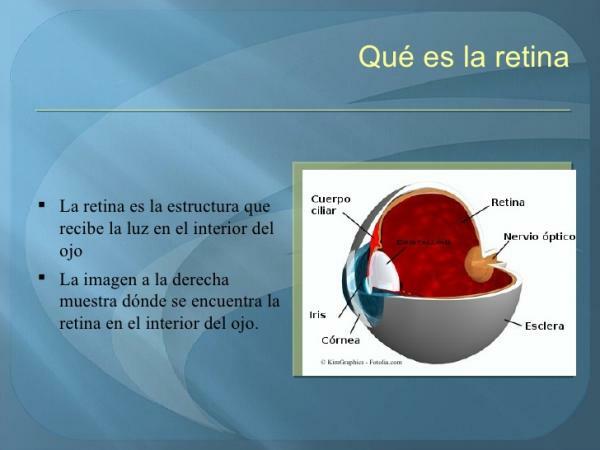
Image: Slideshare
The optic nerve.
The optic nerve is a sensitive nerve, formed by a set of fibers that transmit light impulses from the retina to the cerebral optic chiasm. From this point the visual information is sent to other areas of the brain in the form of electrical signals.
If you want to read more articles similar to Human eye anatomy, we recommend that you enter our category of biology.
References
Did you like this article? Leave your feedback! Do you have any input or questions? Leave your comment in the comment section!