CYTOSKELETON function and structure

In this lesson from a Teacher we will tell you what is the cytoskeleton, what is its function and how is its structure. To begin with, we will focus on the etymology of the word "cytoskeleton": it comes from the Greek "kytos" (nut, cell) and "skeletons" (skeleton, skeletal system, internal bones), its meaning is "proteins that give cells their shape". We can then draw an analogy between the internal skeleton of vertebrates and the cytoskeleton of cells, since in both cases they give shape and structure to the internal parts of a system.
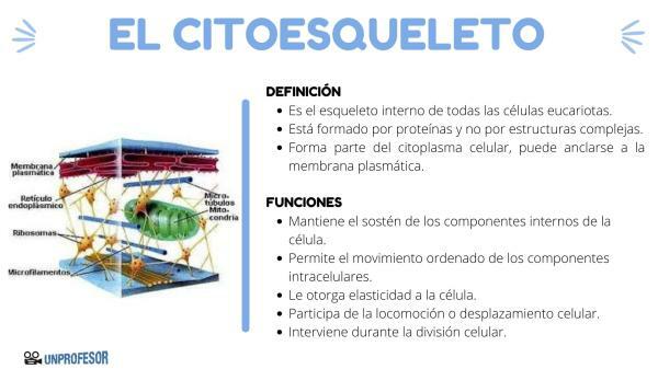
The cytoskeleton is he internal skeleton all eukaryotic cells. We must measure that most cells are microscopic in size, for this reason the cytoskeleton is made up of proteins and not by complex structures like our bones and muscles, even so, they share characteristics such as some of their construction materials and some of their functions.
The cytoskeleton is part of the cell cytoplasm. can be anchored to the plasma membrane. It is made up of fibrillar proteins and resembles an intricate network that allows the movements of the organelles and substances that are within it. the cell, also gives the cell the ability to take various shapes, maintain its volume, and in some cases provides displacement cell phone.
Let's take a closer look at the function and structure of the cytoskeleton.
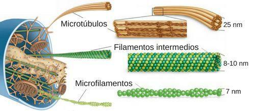
The cytoskeleton is structurally composed of microfilaments or actin filaments, intermediate filaments, microtubules, and the microtrabecular meshwork.
The cytoskeleton structureor is the following.
microfilaments
Is it so made up of a type of protein called actin They are the thinnest and most flexible filaments of the cytoskeleton. They can interact with myosin and other proteins in the cytoplasm and the plasma membrane. Each strand is made up of two intertwined helical chains, we can imagine them as two pearl necklaces that wind around each other.
They have the ability to lengthen or shorten, adding actin units to their ends or removing them, these processes are called polymerization or depolymerization, respectively. This capacity makes the filaments susceptible to deformation and restructuring, in turn influencing cell shape and movement. In interaction with myosin, they are the responsible for the movement of contraction and relaxation of muscle cells.
intermediate filaments
They are named like this, since their size is between the microfilaments that are the smallest and the microtubules that are the largest. They have less elasticity than microfilaments, but offer greater resistance. Like the microfilamentsThey have the ability to arm and disarm. They form a heterogeneous group of diverse proteins.
In epithelial cells, for example, these filaments are made of keratin. In cells of connective tissue, muscle and support cells of the nervous system they are usually vimentin. Neurofilaments belong to neurons. As usual, They provide resistance to mechanical stress and help maintain cell shape. These structures are the most stable of the cytoskeleton and the most insoluble.
microtubules
They are cylinder-shaped filaments or tubes formed by the tubulin protein. They are larger and more rigid than the rest of the structures of the cytoskeleton. Just like the other filaments, microtubules also have the ability to polymerize or depolymerize.
They participate in various functions such as cell transport (movement of vesicles and organelles through the cytoplasm), the movement of cilia and flagella of the cell and, participate in cell division, allowing the movement of chromosomes in order to distribute the genetic material among the daughter cells. Microtubules originate from a structure called centrosome, located near the cell nucleus. This structure is composed of two cylinders called centrioles, Also made up of microtubules.
microtrabecular meshwork
Is a fine and complex three-dimensional network located in the matrix of the cytoplasm that is holding and connecting the different organelles of the cell. Its units are called microtrabeculae and form a mesh that extends throughout the cytoplasm to the cell membrane.

Image: Meanings.com


