Paracentral lobe: characteristics, location and functions
The cortex of the human brain contains several gyri and gyri that delimit different regions. and brain structures, each of them with their respective functions and interconnected with each other. others. One of them is the so-called paracentral lobe, a gyrus located in the medial part of the cerebral hemispheres containing various areas related to planning and managing actions motor.
In this article we explain what the paracentral lobe is, where it is located, what functions the areas that belong to this gyrus perform, and what kind of disorders can be caused if this region of the brain is damaged.
- Related article: "Parts of the human brain (and functions)"
Paracentral lobe: definition and neuroanatomical location
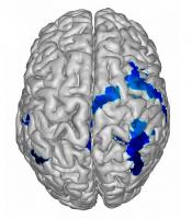
The paracentral lobe is a gyrus of the cerebrum located on the medial surface of the hemisphere, contiguous with the precentral and postcentral gyri. Includes areas of the frontal lobe and parietal lobe. It forms the most medial part of the superior frontal gyrus.
This cerebral region delimits, posteriorly, with the marginal groove; the ascending terminal extension of the cingulate groove, which separates the paracentral lobe from the precunae or precuneus. Its lower limit is the cingulate sulcus, which separates this lobe from the cingulate gyrus. For its part, the central sulcus extends towards the posterosuperior zone of the paracentral lobe, creating the division between the anterior part of the frontal lobe and the posterior part of the lobe parietal.
The cerebrum contains numerous gyri or gyri throughout the entire cerebral cortex, giving it a wrinkled appearance. The cortex is precisely where the higher cognitive functions that involve planning and managing movements or executive decisions are processed and carried out.
The paracentral lobe can be divided into its anterior and posterior portion.: the anterior part of the paracentral lobe is part of the frontal lobe and is often called the supplementary motor area; and the posterior portion is considered as part of the parietal lobe, responsible for the somatosensory functions of the distal extremities. Next we will see what are the main functions of the areas that are included in this part of the brain.
functions
The paracentral lobe is made up of neuronal nuclei that are responsible for motor and sensory innervation of the contralateral lower extremities, as well as the regulation of basic physiological functions, such as urination and defecation.
One of the areas included in this lobe is the supplementary motor area., a brain region that is part of the motor cortex and whose main function is to regulate the production of voluntary movements in the musculoskeletal system. This area, along with the premotor area, both form part of the secondary motor cortex, responsible for planning and initiation of movements that, later, will be in charge of executing the motor cortex primary.
The primary motor cortex, located in the precentral gyrus and paracentral lobe, are somatotopically organized; This means that the different parts of the body that perform movements are overrepresented on a topographic map. precise, such as the hands and face, compared to other areas, such as the trunk and legs, which make movements more thick.
For example, when electrodes are used to stimulate the anterior paracentral lobe, movements of the contralateral leg are initiated. And if these electrodes then move from the dorsomedial to a ventrolateral part in the precentral gyrus, the movements generated will progress from the torso, arm and hand, until reaching the most lateral part of the expensive.
- You may be interested in: "Motor cortex of the brain: parts, location and functions"
Disorders related to damage to this brain region
The main clinical manifestations caused by damage to the paracentral lobe areas usually include motor deficits. Patients may present clinical signs such as paresis (sensation of weakness in one or several muscles) or, directly, plegia or complete muscle paralysis.
Lesions in premotor areas cause alterations in the planning and sequencing of motor actions. Sometimes an impairment or inability to execute learned motor plans is observed, without there being muscle paralysis: a disorder called apraxia.
There are several types of apraxia, but the most common motor syndrome when there is damage to premotor areas usually includes incapacity to use everyday objects and to produce movements with a certain complexity: for example, brushing teeth, opening a door or get dressed. When motor difficulties affect a person's ability to write, the disorder is called agraphia.
Another of the disorders caused by the lesion or resection of the supplementary motor area, located, as we have commented, in the paracentral lobe, is a syndrome that bears his name. Supplementary motor area syndrome affects the ability to initiate movement, initially causing global akinesia. Language disorders may also appear and, later, coordination problems, facial paralysis and hemiplegia contralateral to the damage in this brain region.
In particular, damage to the left supplementary motor area can lead to transcortical motor aphasia, a disorder that causes a lack of verbal fluency, despite the fact that repetition is preserved. There is also a lack of initiative and motivation when establishing communication, and dysnomia may appear (inability to name objects or people) and a slowing of speech, with the appearance of telegraphic language and, sometimes, echolalia (involuntary repetition of words or phrases just heard).
In the most extreme cases, absolute mutism can occur. that prevents the patient from speaking or communicating with others. Motor problems are also relevant, with the appearance of akinesia and loss of movement in the proximal limbs. Difficulties when executing automated movements are also common, although if patients are able to move voluntarily they do not usually present these alterations.
Bibliographic references:
- Cervio, A.; Espeche, M.; Mormandi, R.; Alcorta, S.C. & Salvat, S. (2007). Postoperative supplementary motor area syndrome. Report of a case. Argentine Journal of Neurosurgery, 21 (3). Autonomous City of Buenos Aires.
- Roland, P. E., Larsen, B., Lassen, N. A., & Skinhoj, E. (1980). Supplementary motor area and other cortical areas in organization of voluntary movements in man. Journal of neurophysiology, 43(1), 118-136.
- Snell, R. S. (2007). clinical neuroanatomy. Pan American Medical Ed.