Leg muscles: types, location, characteristics and functions
The locomotor system is made up of the osteoarticular system (bones and joints) and the skeletal muscles of the body, that is, those that respond to voluntary actions.. To give you an idea of the biomechanical work of art that we have in our body, it is enough to say that we have 206 bones, 360 joints (86 of them in the skull) and more than 600 skeletal muscles, all of them with a common mission: that we can maintain our shape and move.
The voluntary musculature (skeletal or striated) makes up approximately 40% of the weight of an adult man, so its functionality is counted for itself. If we look at the legs, the third lower pelvic segment between the knee and the ankle, it is estimated that a human being walks about 3,500 million kilometers throughout his life. For this reason, the leg muscles must be prepared for a slight but continuous exercise throughout our existence.
For all these reasons, it is also common to see how overweight and obese people have serious problems moving around in the lower body. An added body kilogram is equivalent to 7 more in the knee portion, so increasing 10 kilos of mass, theoretically, is associated with 70 kilograms more pressure on this joint.
With all this data in hand, it is more than clear to us that the leg is an essential anatomical section of the human being, both for locomotion and for supporting our weight in an environment three dimensional. In honor of these structures, today We present you all the muscles of the legs and their particularities.
- Related article: "Locomotor system: what it is, parts and characteristics"
leg muscles
As we have said, the term "leg" refers to the lower extremity of the human body, which runs from the trunk to the foot. In some contexts it is considered to also include the foot, and in others the thigh is excluded. In any case, we are going to perceive the structure as a "whole" at the anatomical level, ranging from the base to the tips of the fingers. We start by naming the muscles in the anterior compartment of the leg.
1. Muscles in the anterior compartment of the leg
Here we find 4 well-differentiated muscle groups: tibialis anterior, extensor digitorum longus muscle, extensor hallucis longus muscle (extensor hallucis) and third peroneus muscle. We tell you its peculiarities.
1.1 Tibialis anterior
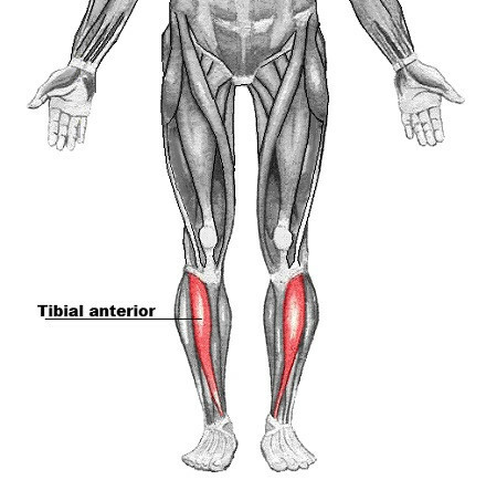
The anterior tibial, as its name suggests, accompanies the tibia on its lateral surface.. The contraction of the tibialis anterior stabilizes the ankle, especially at the moment in which the sole of the foot makes contact with the surface of the ground in the action of walking.
In general, its function is to keep the leg stable while walking, regardless of the condition of the terrain or its inclination.
1.2. Extensor digitorum longus (EDL) muscle
We are dealing with a penniform muscle (shaped similar to that of a feather) innervated by the deep peroneus. It is an important dorsiflexor and therefore has the function of producing the simultaneous extension of almost all the toes.except for the fat one.
It originates from the lateral condyle of the tibia and the medial surface of the fibula. The fibers of this musculature come together in a tendon, which travels along the dorsal surface of the foot, to be divided into 4 units that are inserted into each toe.

- You may be interested in: "Muscular system: what it is, parts and functions"
1.3. extensor muscle of the hallucis
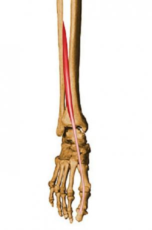
As you can imagine, this is the muscle responsible for extension of the big toe, but also for dorsiflexion of the sole of the foot (together with the extensor digitorum longus). It originates on the medial surface of the fibula shaft and inserts on the phalanx of the great toe..
1.4. third peroneus muscle
This is a small muscular belly, located between the anterior and external portion of the leg. It originates with the EDL on the medial surface of the fibula, and runs with it for a certain distance, until it reaches the dorsal surface of the foot. Here, the third peroneus muscle is divided and attached to the fifth metatarsal. Along with other muscles, it is responsible for flexion and eversion of the foot..

2. Muscles in the lateral compartment of the leg
We continue on our journey, this time on the side of the leg, paying attention to the following muscle groups: the peroneus longus and the peroneus brevis. We go with them.
2.1. peroneus longus
This muscle is located on the lateral and outer surface of the leg. It originates from the lateral aspect of the fibula and tibial condyle, fusing along its course into a tendon that inserts on the medial cuneiform and the base of metatarsal I. Its main function is to extend the foot on the leg, take it out and allow it to perform a rotation movement..

2.2. peroneus brevis
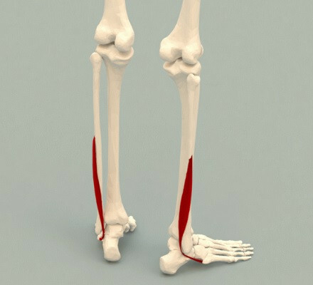
Located on the outer side of the leg and below the knee, this muscle is responsible for allowing eversion of the foot. It inserts on the anterolateral surface of the fibular shaft and anchors on a tubercle associated with metatarsal V..
- You may be interested in: "Muscle fiber: what it is, parts and functions"
3. Muscles in the posterior compartment of the leg
We expedite the process, because in this last section, a total of 7 muscles await us that must be described, at less superficially, which in turn are organized into two faces (superficial and deep, divided by a fascia). Let's go there.
3.1. gastrocnemius
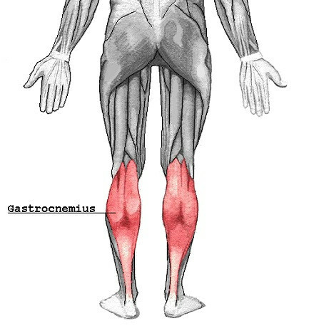
We are entering familiar territory, as this muscle, divided into two halves, is what we popularly know as “the twins”. First of all, it should be noted that it has two "heads", one medial and one lateral, which converge in the ventral section. It is located on the soleus muscle and occupies a large part of the posterior aspect of the leg, from the knee to the ankle.. It is the main motor of the beginning of the physical march.
3.2. Plant
The plantar muscle lies deeper than the gastrocnemius and is much smaller in size, with a narrow diameter. It is absent in 10% of the world population and its function is very weak., so we are not going to dwell on it anymore.

3.3. soleus
As we have said before, it is located below and behind the gastrocnemius. Curiously, it is called soleus because of its flattened and circular shape, which makes it look like a sole.

3.4. Popliteal
The muscles that we have named so far (gastrocnemius, plantaris, and soleus) form the external face of the posterior compartment of the leg. As of now, the following muscle groups are located in the deep section of it.
The popliteus is located at the top of the leg, "above the knee". It is located anterior to the gastrocnemius and is short, flattened, and triangular in shape. Its function is to laterally rotate the femur on the tibia, thus allowing flexion of the knee joint.

3.5. posterior tibial
elongated in shape, This muscle is located between the flexor digitorum longus and the flexor hallucis, old acquaintances that we have already addressed. Its function is to allow adduction of the foot, stabilize the plantar arch and allow plantar flexion of the foot.
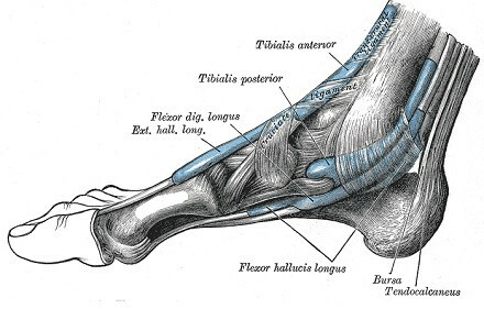
3.6. flexor digitorum longus muscle
This muscle originates from the medial part of the posterior aspect of the tibia. While the extensor digitorum longus (EDL) muscle was responsible for the extension of the 4 fingers, this is the one that allows its bending.

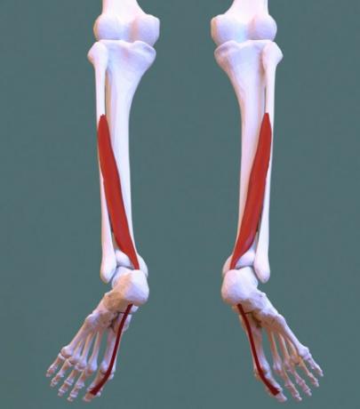
3.7. flexor hallucis longus muscle
The same premise as the previous case, but with the big toe. Simple as that.
Summary
As you will have been able to verify, we have counted a total of 13 muscles: 4 in the anterior compartment, 2 in the lateral compartment and 7 in the posterior compartment. Undoubtedly, the back face is the one that sounds the most familiar to all of us, since here are the twins, the soleus or popliteus, muscle groups that many of us have had to learn during a lesson in biology.
Except for the plantar, all these muscles play an essential role in the functioning of the leg and feet alike. Thanks to them, we are able to move effectively at different speeds and on different terrains.
Bibliographic references:
- Jacobs, R., Bobbert, M. F., & van Ingen Schenau, G. J. (1996). Mechanical output from individual muscles during explosive leg extensions: the role of biarticular muscles. Journal of biomechanics, 29(4), 513-523.
- Joseph, J., & Nightingale, A. (1952). Electromyography of muscles of posture: leg muscles in males. The Journal of physiology, 117(4), 484-491.
- Muscles of the leg, teach me anatomy. Collected on April 11 in https://teachmeanatomy.info/lower-limb/muscles/leg/
- Valentino, B., & Melito, F. (1991). Functional relationships between the muscles of mastication and the muscles of the leg. Surgical and Radiologic Anatomy, 13(1), 33-37.
