Cartilaginous joints: what they are, types and characteristics
The locomotor system refers to the set of tissues and organs that allow living beings to move and respond to environmental stimuli. In the case of humans, this intricate work of biomechanics has 206 bones, more than 650 muscles and 360 joints, of which 86 are located in the skull.
When we talk about the formation of movement in vertebrates, we automatically think of muscles and bones, since they are "the bulk" that causes force when responding to a stimulus. In any case, we must not forget about the joints, since these are the ones that unite two or more bones so that they maintain their structure or, failing that, allowing movement.
Based on this premise, we find it interesting to investigate the world of joints and everything that their anatomy and variability entails. In the following lines, we tell you everything about cartilaginous joints and their particularities.
- Related article: "Locomotor system: what it is, parts and characteristics"
What is a cartilaginous joint?
As indicated in the medical dictionary of the Clínica Navarra University (CUN),
A joint is a specialized structure in the area of union between two or more bones or, failing that, between bone and cartilage structures.. Normally, when we think of joints, the knee and elbow come to mind, but these obvious formations are in the minority.Without going any further, 86 joints are found in the head, of which the head of the atlanto-occipital and the temporomandibular joint are the only mobile ones. In the skull, some of the joints are nothing more than the "glue" between the flat bones that protect our brain, but how they use the classic definition (union of two or more bones), they fall into this great category anatomical.
Within the grouping of joints, we find three large categories. These are the following:
- Synovial joints: the bones involved are separated by a very narrow joint cavity, which allows a wide range of motion. The elbow and knee are clear examples.
- Fibrous joints: connections between two bones attached directly to each other, often by fibrocartilage. They are rigid. An example of this are the points of union between the bones of the head, known as sutures.
- Cartilaginous joints: those that concern us here.
Cartilaginous joints are at a midpoint on a physiological level, as they are more mobile than fibrous but less than synovial, which represent the maximum range of mobility. Furthermore, it should be noted that cartilaginous joints also form the growth regions of the long bones and intervertebral discs of the human spine.
Types of cartilaginous joints
Cartilaginous joints include the symphyses and synchondrosis. We tell you about its particularities in the following lines.
1. synchondrosis
In synchondrosis, the connecting element between the bones involved is the hyaline cartilage, as opposed to the fibrocartilage of the symphyses. (although some symphyses also have hyaline cartilage). In addition, this time the training is transitory.
An example of this is the joint present between the basilar process of the occiput and the body of the sphenoid, when both structures are still cartilaginous because they have not completed their development. Once the relevant tissue maturation occurs, both articular surfaces fuse and the synchondrosis disappears. They usually appear between bony growth structures allowing a certain degree of movement, but they ossify completely with time.
On the other hand, it should also be noted that there is a pair of permanent synchondroses. One of these is the first sternocostal joint, where the first rib and the manubrium of the sternum meet. This stands out above the rest, since the other rib-sternum junctions are of the flat synovial type. The other permanent synchondrosis is the petro-occipital, between the occipital and petrous bones of the skull.

Synchondrose between long growth bones
In the long bones of humans (such as the femur) two very specific structures are distinguished: the epiphysis and the diaphysis.. The epiphysis is each of the two ends of the long bone, the area where the joints are located, with a greater width than the diaphysis. For its part, the diaphysis is the area between the two epiphyses, which is covered with a hard periosteum and in its internal zone contains the bone marrow, where the circulating cellular elements (red blood cells and others).
Synchondroses are usually located in the growing long bones between both epiphyses and the central diaphysis. These relatively "soft" joints allow the body of the bone to elongate and separate the conglomerate. bone into three distinguishable sections, as if they were three bones (epiphysis-cartilage-diaphysis-cartilage-epiphysis). Ultimately, these cartilages ossify and form an anatomical whole.
- You may be interested in: "Diarthrosis: what they are, types and anatomical characteristics"
2. symphysis
In this type of cartilaginous joint, the bones in contact are first united by a fibrocartilaginous nature sheet (fibrocartilage), which fuses the components into an anatomical structure. Unlike the synchondrosis that we will see later, the symphyses are permanent throughout the life of the individual.
A clear example of a symphysis is the pubic symphysis, although there is also one present in the mandible, in the sacrococcygeal region, in the sternum and, without going any further, between the vertebrae of the spine. Simply put, in the symphysis two separate bones are connected to each other by cartilage.
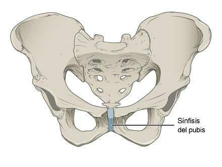
symphysis pubis
The pubic symphysis (the most conspicuous of all) is defined as a cartilaginous joint located at the confluence of the two pubic bones, formed by a small fibrocartilaginous disc that is interposed between two narrow sheets of hyaline cartilage. In addition, this disc is reinforced by a particularly interesting pair of ligaments: the inferior and superior pubic ligaments. These provide enormous stability to the osteoarticular conglomerate of the pubis.
Curiously, the female pubic symphysis is covered by adipose tissue, which forms the well-known "mount of Venus". The characteristic female pubic hair grows on this structure, but it also has glands that secrete hormones involved in sexual attraction. Even the most seemingly anecdotal anatomical variation has clear evolutionary significance.
Cartilaginous joints and the vertebral column
As we have said, the intervertebral discs are fibrocartilaginous symphyses, which are located between each of the 26 vertebral bones that provide axial support to the trunk and provide protection to the spinal cord, which allows the transmission of information to our endings nervous.
Have you ever wondered why the elderly seem to shrink as they get older, right? Interestingly, much of this shrinking is due to the degradation of the intervertebral joints, in addition to osteoporosis and other processes of bone damage and remodeling. The force of gravity acts on the spine and, over the years, the vertebrae compress these discs and squeeze each other.
After 40 years of age, people usually lose one centimeter in height for every 10 years, partly due to this compression caused by the wear that occurs in the intervertebral discs.: we remember again that they are cartilaginous joints of the symphysis type. As they age, an average human being can lose from 2 to 7.5 centimeters in height throughout their lives, a value that seems insignificant but is more than remarkable.
Osteoporosis is also essential to understand this reduction in height, because in it, bones are resorbed and destroyed at a higher rate than their synthesis. As a result, some long and short bones thin and shorten further over time, becoming much more brittle and prone to fracture. It is no coincidence that almost no one presents a vertebral fracture before the age of 50.
Summary
As you may have observed, cartilaginous joints go far beyond the merely anecdotal, because thanks to they, for example, support the vertebral column and can explain much of the reduction in height in humans Adults. On the other hand, thanks to the cartilaginous articulation of the pubis, the mount of Venus can be manifested in women, which seems to play a not insignificant role in sexual attraction.
With these lines, it is more than clear to us that, in the human body, almost everything has a reason. Except for a few vestigial structures (such as wisdom teeth), every tissue, cell, and junction point presents a specific function, more or less important to maintain internal homeostasis or perform movements in the environment.
Bibliographic references:
- Why do people shrink? radyschildren.org. Collected on March 28 in https://www.rchsd.org/health-articles/por-qu-se-encoje-la-gente/
- Anatomy Notes. Types of joints: synovial and solid, Elsevier. Collected on March 28 in https://www.elsevier.com/es-es/connect/medicina/anatomia-tipos-articulaciones-sinoviales-y-solidas.
- Articulation, Navarra University Clinic (CUN). Collected on March 28 in https://www.cun.es/diccionario-medico/terminos/articulacion.
- Cartilage joints, lumenlearning.com. Collected on March 28 in https://courses.lumenlearning.com/boundless-ap/chapter/cartilaginous-joints/.
- Cartilagous joints, radiopaedia. Collected on March 28 in https://radiopaedia.org/articles/cartilaginous-joints.
- Montero, S. TO. R. (2007). Pubic symphysitis. Bibliographic review. Seminars of the Spanish Rheumatology Foundation, 8(3), 145-153.