All shoulder JOINTS
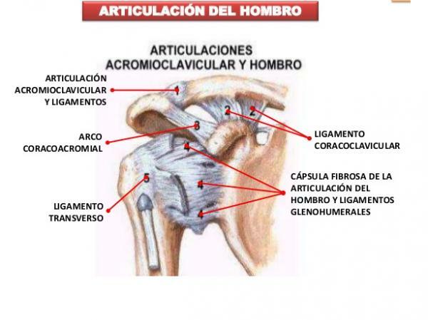
Image: pc13sport.com
The shoulder is the part of the human anatomy that unites each of the arms with the trunk. Although the shoulder is not simply an area of attachment. The bones, muscles, ligaments and tendons that form it allow us to perform different types of arm movements (extension, flexion, abduction, adduction and internal rotation and external). In this lesson from a TEACHER we will review what are the joints of the manor, its characteristics and by what bones the shoulder joints are formed. If you want to know more, we invite you to continue reading!
Index
- The shoulder joint complex
- Scapulohumeral or glenohumeral joint
- Sternocostoclavicular joint
- Acromioclavicular joint
- Subdeltoid or suprahumeral joint
- Scapulothoracic joint
The articular complex of the shoulder.
The shoulder is commonly believed to be a joint, but the reality is somewhat different. The shoulder is actually a complex or set of joints: 5 joints to be specific. The shoulder joints are formed by the union of four bones: the humerus (forearm bone), the clavicle, the scapula or shoulder blade, and the sternum. If you want to know more about these bones you can consult the lesson Bones of the shoulder.
The four bones that make up the shoulder join or articulate, giving rise to five different joints. Next we will see them, one by one.
In this other lesson we will discover all the joints of the human body.
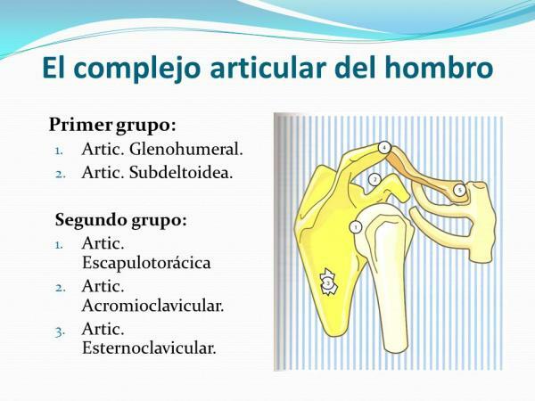
Image: SlidePlayer
Scapulohumeral or glenohumeral joint.
The glonohumeral joint is formed by the union of the humerus head (end of arm bone) and the glenoid cavity. The glenoid cavity is a depression that is located on the sides of each of the clavicles and is where the head of the humerus is located or housed.
This type of union in such peculiar cavity allows one of the shoulder joints to be with more mobility. This joint can move in three planes of space and allows flexion, extension, abduction, adduction and rotation movements as well as providing stability. Most of these movements involve the rotation and friction of bones, so it is important that the ligaments and muscles involved in the joint are in good condition to prevent inconvenience.
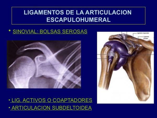
Image: Slideshare
Sternocostoclavicular joint.
The sternocostoclavicular joint, as its name indicates, is the one in which the union of each of the sides of the sternum to each of the clavicles. In addition, the first costal cartilage is also attached to this joint, which is responsible for attaching the first rib to the sternum.
The movement of this joint is more limited and it only takes place in two planes of space. In summary, movement in these two planes of space allows the clavicle to move vertically or anteroposterioment, although the movement is very limited. In addition, a slight rotational movement can be produced thanks to a fibrocartilage disc that separates the two parts of the joint: the sternum-clavicle joint and the joint sternum-rib.
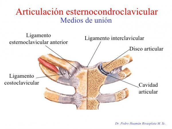
Image: friends forever
Acromioclavicular joint.
The acromioclavicular joint is made up of the junction of the acromion and the lateral end of the clavicle. The acromion is a protrusion on the outside of the scapula that goes towards the upper and anterior part of the arm. This structure, which can be more or less large or flat depending on the person, joins the clavicle.
As in the previous case, between the two bones there is a fibrocartilage disc and it is strongly reinforced by ligaments and muscles. This joint makes it possible to reinforce the movements made by the clavicle and mainly allows us to raise our arms above our head.
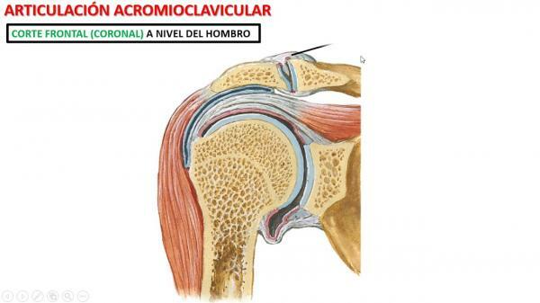
Image: YouTube
Subdeltoid or suprahumeral joint.
If you consult other sources, you will see that the subdeltoid joint does not always appear. This is because the subdeltoid joint is sometimes not named within the shoulder joints because it is a false joint. This joint is of the synsarcosis type, that is, instead of joining two bones, joins two muscles. In this case, the subdeltoid joint allows gliding of the deltoid and supraspinatus.
The function of the subdeltoid joint is help perform their movements to the scapulohumeral joint since both are mechanically united. For this sliding movement between the two muscles to take place, between the two there is a bag of special fluid (synovial fluid) that serves as a lubricant and prevents chafing and deterioration of the muscles. It is important that the liquid inside this bag remains with the appropriate composition and in good condition, since if no discomfort may appear during movements.
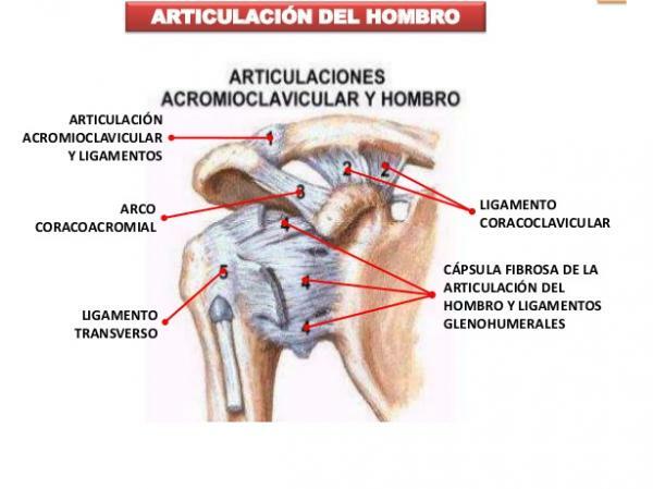
Image: Slideshare
Scapulothoracic joint.
The scapulothoracic joint is the one in which the junction of the scapula with the chest wall, mainly with the ribs. These two structures are covered by two muscles: the subscapularis muscle in the case of the scapula and the serratus anterior muscle in the case of the chest wall. Actually, the joint occurs between these two muscles when moving one over the other, so as in the previous case we are dealing with a case of a sisarcosis-type joint. Furthermore, as in the previous case, we are faced with a false joint since it is mechanically joined to another, the acromioclavicular joint.
Despite being a false joint, the scapulothoracic joint is one of the shoulder joints more important since it will allow us to have a great range of motion of the arms. Very important and powerful muscles such as the serratus muscles (serratus major and minor) or the trapezius are related to this joint.
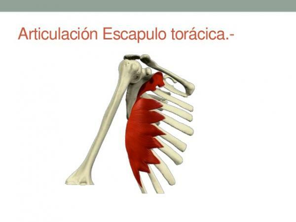
Image: Slideshare
If you want to read more articles similar to Shoulder joints, we recommend that you enter our category of biology.
Bibliography
- Physiosaludable (May 3, 2017). How many joints make up the shoulder. Recovered from https://www.fisiosaludable.com/
- Biomechanical Rehabilitation Medicine (s.f). Joint complex of the shoulder. Recovered from http://www.sld.cu/sitios/rehabilitacion-bio/temas.php? idv = 18657
Dolopedia (April 2019). Shoulder joints. Recovered from https://dolopedia.com/categoria/articulaciones-del-hombro

