Discover the TYPES of VERTEBRAS of the human body
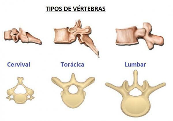
Image: Natural Sciences
The vertebrae are the bones that make up the spine, the central axis of the axial skeleton of more than 50,000 animals that have this structure. The vertebrae are extremely irregular bones, they have different shapes according to what we refer to: the vertebrae of the neck or beginning of the back are quite different from the vertebrae that make up the end parts of the spine vertebral. In this lesson from a TEACHER we will see what vertebrae are and what their parts are, as well as the types of vertebrae that make up the spine of humans.
Index
- The vertebrae and their parts
- Cervical vertebrae, one of the types of vertebrae
- Thoracic vertebrae
- Lumbar vertebrae, another type of vertebra
- Sacrum and coccyx
The vertebrae and their parts.
Before knowing the types of vertebrae that exist, we are going to discover the different parts of this area of the human body. The vertebrae They are irregular bones, made up of bone tissue and hyaline cartilage.
Although the vertebrae are different depending on the region of the spine to which they belong (thoracic, lumbar, etc.), there is a basic model of vertebra in which we can distinguish the following parts:- Body. The body of the vertebra is the central region of the vertebra and has a cylindrical shape; allows each of the vertebrae to bear its proper weight.
- Articular processes. They are each of the projections that each of the vertebrae have to articulate with each other. A typical vertebra has four processes: two ascending or superior and two descending or inferior. The processes are located on the sides of the foramen or vertebral canal.
- Transverse processes. Like the previous ones, they are projections but in this case they are on the sides of the vertebra.
- Spinous apophysis. The spinous processes are found in the posterior part of the vertebrae, incorporated into it by the lamina.
- Vertebral laminae. The vertebral laminae have this shape because they are laminates of bone, more or less square and flat, which form most of the posteolateral part of the spinal foramen. Each of the vertebrae has two vertebral plates, one on the right and one on the left.
- Medullary canal. It is located in the center of the vertebra and the spinal cord runs through the spinal canal made up of all the vertebrae.
In the spinal column we can distinguish three regions: cervical region, thoracic region and lumbar region. Each of the vertebrae in these regions have characteristics and, within them, some of the vertebrae have very particular characteristics. If you want to know more about the characteristics of each of the vertebrae, keep reading!
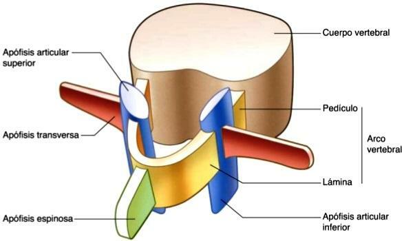
Image: Spinal column
Cervical vertebrae, one of the types of vertebrae.
We humans have seven cervical vertebrae, of which two of them are so different from the rest that they have common names: atlas and axis. All cervical vertebrae are named with a capital letter C followed by the number they occupy, starting from the head; for example, the first vertebra is called C1 or atlas while the last, which articulates with the first thoracic vertebra, is called C7.
The cervical vertebrae are smaller and more fragile than those of the rest of the spinal column. In addition, its vertebral canal is triangular in shape, its spinous process has two protrusions (spinous process bifida) and has transverse holes that allow the passage of the vertebral artery and vein and the nerves friendly. The first two cervical vertebrae have characteristics that differentiate them so much from the rest that they have their own names, they are called atlas and axis, respectively.
- C1 is also called atlas, as in Greek titan that supports the sky on its shoulders since it is responsible for supporting the head through the occipital bone. A key feature of the atlas is that it lacks a vertebral body and spinous process. The vertebral body is replaced by two lateral masses, which articulate with the occipital bone and the axis, and which are joined by an anterior and a posterior arch.
- C2 is also called axis. This vertebra has an odontoid process (also called dens) that extends across the top of the vertebra, from the front of the vertebra. Its peculiar articulation with the anterior vertebra, atlas, makes it possible to move and rotate the head independently of the torso.
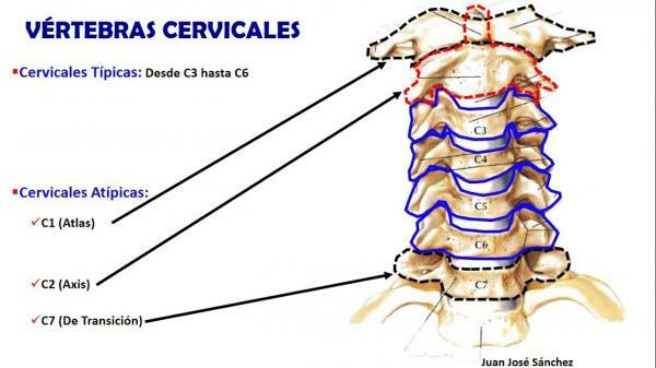
Image: Youtube
Thoracic vertebrae.
Another type of vertebrae are thoracic and human beings have 12 that are named as T, followed by a number ranging from 1 to 12. The thoracic vertebrae are easily identified by having, on the sides, the costal fossae, three regions (upper, lower, transverse) that are loosely joined with the head of the ribs. In addition, the shape of the vertebral body of these vertebrae is slightly reminiscent of a heart and its medullary canal is more rounded than in the previous ones.
The size of the thoracic vertebrae is greater than that of the cervical vertebrae, and it increases as we go down the back. Finally, in the thoracic vertebrae they can be more easily identified intervertebral discs; intervertebral discs are disc-shaped structures composed of a gelatinous nucleus surrounded by a fibrocartilage ring whose function is to cushion the movement of the vertebrae and reduce friction between they.
Of all of them, the T1 vertebra is the most different since it has a complete articular fact for the insertion of the first rib and a face for the upper half of the head of the second rib. In addition, it has the spinous and transverse processes much longer than the rest of the vertebrae in this area.
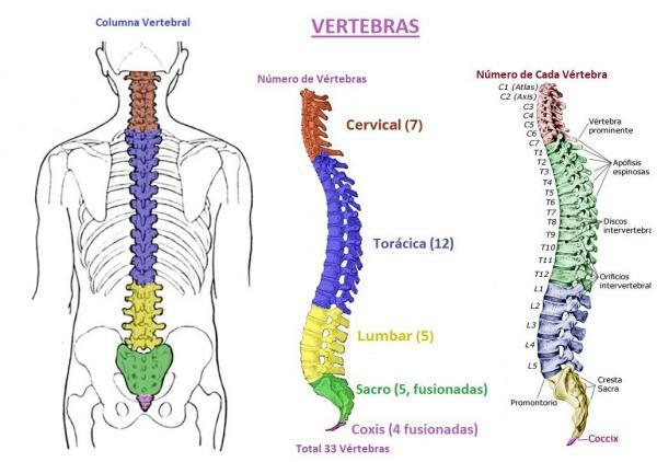
Image: Natural Sciences
Lumbar vertebrae, another type of vertebrae.
The lumbar vertebraeIn humans, they are the largest and strongest of the entire spinal column since they have to support more weight than the rest. Humans have 5 lumbar vertebrae, some of which have undergone great modifications throughout human evolution.
In addition to their large size, the lumbar vertebrae stand out for their kidney-shaped body, their spinal canal in the shape triangle and its long and thin processes, which together with the irregular shape of the intervertebral discs allow a greater flexion and extension of the spine.
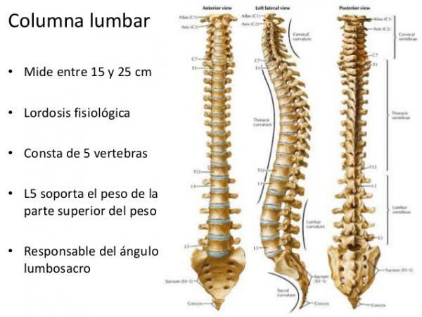
Image: Slideshare
Sacrum and coccyx.
At the end of the spine we find two bone complexes: the sacrum and the coccyx or coccyx. In humans, the sacrum is formed by the fusion of five vertebrae while the coccyx is formed by the fusion of three to five vertebrae.
This fusion begins during embryonic development and ends during infancy. Being fused, these bone complexes lack joints.

Image: Medline Plus
If you want to read more articles similar to The types of vertebrae, we recommend that you enter our category of biology.
Bibliography
- Wikipedia (May 28, 2019). Vertebra. Recovered from https://en.wikipedia.org/wiki/Vertebra
- Educational magazine Partsdel.com. (May 2017). Parts of the vertebra. PartsDel.com. Recovered from https://www.partesdel.com/vertebra.html.
- Topographic anatomy (s.f) Cervical vertebrae Recovered from https://www.anatomiatopografica.com/huesos/vertebras-cervicales/
- Topographic anatomy (s.f) Thoracic vertebrae. Recovered from https://www.anatomiatopografica.com/huesos/vertebras-toracicas/
- Topographic anatomy (s.f) Lumbar vertebrae. Recovered from https://www.anatomiatopografica.com/huesos/vertebras-lumbares/
- Topographic anatomy (s.f) Sacral bone. Recovered from https://www.anatomiatopografica.com/huesos/hueso-sacro/
- Topographic anatomy (s.f) Coccyx bone. Recovered from https://www.anatomiatopografica.com/huesos/hueso-coccix/


