Structure of the BONES of the human body
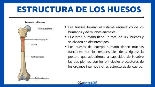
The bones They are the fundamental pillars that support our body, but their complexity goes far beyond what we can see with the naked eye. These structures are made up of a complicated network of bone tissue, specialized cells and minerals that work together to provide strength, flexibility and support to our musculoskeletal system.
In this lesson from a TEACHER, we are going to explain to you what is the structure of bones and we will thoroughly analyze the internal architecture of this system of our body.
Index
- What are bones
- What is the structure of bones
- How bones grow
- bone diseases
What are bones?
The bones form the skeletal system of humans and many animals. The human body has a total of 206 bones and they are divided into different types, which are classified according to their general characteristics, such as their shape, location, properties, etc. We can differentiate between 5 different types of bone:
- Long bones: They are those that have a length greater than their width and thickness. They are dense and strong bones in which the red and yellow marrow is found.
- Short bones: These are bones that have similar measurements in length, width and thickness.
- Flat bones: A good length predominates in these bones, but the width and thickness are quite scarce. That is because it is usually part of the framework of different cavities in the body.
- Irregular bones: In this last category are all bones that have an irregular shape and that prevents them from being part of any previous classification.
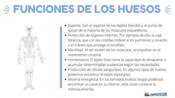
The bones of the human body have many important functions. First of all, they are responsible for the rigidity of the structure of our body, the contour of the structure, the posture we acquire, the ability to walk on both legs and movement. Secondly, thanks to their rigidity, bones are the main protectors of internal organs and other structures of the body.
Here we discover the functions of human bones and his composition.

What is the structure of bones.
The bones are made up of three servings:
- Diaphysis: It is the central part of the bone.
- Epiphysis: They are the ends that we find in long bones.
- Metaphysis: It is the intermediate portion of the bone.
Beyond this general structure, bones are made up of several layers and each of them has a specific function. We are going to explain to you all the parts of the bone structure, starting from the most internal, to the most external.
Medullary cavity
This is one hollow region in the bones, where is the bone marrow, which is responsible for producing red blood cells. Usually, the medullary cavity is located in the diaphysis of the bones.
Endosteum
The endosteum It is a very thin membrane of connective tissue found surrounding the interior of the medullary cavity of the long bones.
nutrient artery
The nutritive artery is in charge of provide blood to the bone, through the nutritional holes. This is then distributed throughout the bone through capillaries that are increasingly thinner. The blood provides the nutrients and oxygen necessary for the proper functioning of the bone.
Woven bone
He woven bone It is the main component of bone and is made up of several types of bone cells (osteocytes, osteoblasts, osteoclasts and stem cells). 2% is bone tissue, while 70% is resistant extracellular substance (hydroxyapatite). They themselves secrete this extracellular substance, from calcium and phosphorus. 30% belongs to collagen.
Periosteum
The periosteum It is the membrane of fibrous and resistant connective tissue that covers the bones in its external region.

How bones grow.
When you stop to think about the skeleton, you might think that the bones are not alive, but rather are like wooden sticks that support us. However, you should keep in mind that bones are living tissues and like all living tissues, they need a constant supply of blood to supply them with oxygen and nutrients.
The children's bones They grow by lengthening from the ends. The ends of children's bones have a soft part called plate or growth cartilage. These plates generate new bone that is added to the previous one and lengthens the bones little by little. During adolescence, children's growth plates turn into hard bone, so the bones stop lengthening.
However, even the bones of adults are constantly enlarging and reforming in a process known as remodeling.
In the remodeling process, Old bone tissue is gradually replaced by new bone tissue. Each bone is completely remodeled every 10 years. For that reason, bones need a constant supply of calcium, minerals, vitamin D and growth hormones.
Bone diseases.
When the structure of the bones does not function correctly and does not work in an organized way to remodel itself, they can some diseases occur:
- Osteoporosis: It occurs when old bone is removed more quickly than new bone, resulting in bone that is weaker and more prone to fracture.
- Osteopetrosis: This disease is the opposite and occurs when bone removal is too slow and the bones become too dense.
- Imperfect osteogenesis: People with this disease have a genetic defect that does not allow the body to generate enough collagen or it is produced incorrectly.
- Paget's disease of bone: This disease refers to people in whom more bone is deposited than is removed and, therefore, new bone does not form correctly.
- Fibrous dysplasia: Finally, we want to talk to you about Fibrous Dysplasia, which is a disease in which normal bone is replaced with a fibrous tissue, similar to a scar, but which is not strong enough to perform the function of the bones.
We hope this lesson has helped you understand a little better. bone structure and the importance of these in our body. If you want to continue delving a little deeper into the anatomy of the human body, do not hesitate to consult our biology section.
If you want to read more articles similar to bone structure, we recommend that you enter our category of biology.
Bibliography
- Arsuaga, J. L., Martınez, I., Gracia, A., Carretero, J. M., Lorenzo, C., García, N., & Ortega, A. YO. (1997). Chasm of bones (Sierra de Atapuerca, Spain). The site. Journal of Human Evolution, 33(2-3), 109-127.
- Ortiz Cruz, E. J., Campo Loarte, J., Martínez Martín, J., & Canosa Sevillano, R. (2000). Structure and organization of a bone and tissue bank. Rev. orthop. traumatol.(Mother, printed ed.), 127-138.

