ORGANS of the circulatory system and their FUNCTIONS [with images]

The circulatory system The cardiovascular system is one of the most important systems of the human body, since it is in charge of carrying out fundamental activities in the life of any organism, such as the transport of oxygen and nutrients to all cells of the body, hormones for communication between organs, cells of the immune system, regulate body temperature or balance water. In this lesson from a PROFESSOR we are going to tell you which are the organs of the circulatory system. Join us to find out more!
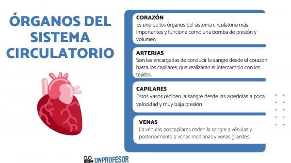
The heart is one of the organs of the circulatory system most important and works as a pressure pump and volume, being able to pump approximately 5 L of blood per minute at rest. The heart is the size of a fist and is located in the upper part of the thorax, slightly to the left side, although there are pathologies where it is located to the right. Inside out they are distinguished three hats:
- Inner layer or endocardium
- Middle layer or myocardium, formed by the heart muscle itself
- Outer layer or epicardium
In turn, the heart is surrounded on the outside by a connective tissue membrane called the pericardium. This membrane holds the heart in position and allows the heart to contract and relax. The pathology of this membrane can become serious, causing, for example, cardiac tamponade.
The heart works by generating a pressure wave that starts in cells known as the cardiac pacemaker and spreads through an electrical conduction circuit that runs through all cavities. Thanks to this pressure wave, a volume is pumped to the four cavities and to the arteries of the body, which lead it to the tissues. Subsequently, the blood re-enters the heart through the veins and the cycle is restarted.
The heart chambers
The heart is organized into four chambers: two atria and two ventricles, divided into left and right. In addition, there is a septum or septum that separates both sides and a fibrous cardiac skeleton that separates the atria from the ventricles. Blood flow flows between heart chambers and in its communication with blood vessels through valves. The four cavities are:
- Right atrium (RA): it is the cavity where the cells are found where the cardiac impulse begins (pacemaker cells) and which is located in a region called the sinoatrial node. This impulse passes through some routes to the atrioventricular node and from there through other routes to the ventricles. After their contraction, they pump the blood that reaches them through the superior and inferior vena cava towards the right ventricle through the right atrioventricular valve or tricuspid valve.
- Right Ventricle (RV): the pressure wave from the right atrium reaches this cavity and contracts. After its contraction, it pumps the blood that comes from the right atrium to the pulmonary artery through the right semilunar valve or pulmonary valve for oxygenation in the lungs.
- Left atrium (LA): the pressure wave reaches this cavity directly from the right atrium through a particular pathway. After its contraction (synchronous to the other atrium), it pumps the oxygenated blood that reaches it through the pulmonary vein into the left ventricle through the left atrioventricular valve or valve mitral.
- Left ventricle (LV): the pressure wave reaches this cavity from the right atrium and contracts, together with the other ventricle. After its contraction, it pumps the blood that reaches it through the left atrium towards the aorta artery through the left semilunar valve or aortic valve. The left ventricle has a very thick layer of heart muscle, as it is the chamber that must develop the greatest pressure to pump blood.
They do not constitute organs of the circulatory system as such, but are rather compliant ducts regards of drive the blood pumped by the heart to all the organs and tissues of the body and back to the heart. There are three general classes of glasses: arteries, veins and capillaries.
Arteries
The arteries They are in charge of conducting the blood from the heart to the capillaries, which will carry out the exchange with the tissues. Through them the blood flows under great pressure, which together with gravity (to a lesser extent), are the driving forces of the blood. There are three classes of artery depending on their size
- Large arteries: they leave directly from the heart and are the aortic artery and the pulmonary artery. The aortic artery directs blood to the major or systemic circuit and the pulmonary artery to the lungs for gas exchange. The blood from the aortic artery is pumped to a very high pressure, while the pulmonary to a lower pressure, and in both cases varies between a maximum (systolic pressure) and a minimum (pressure diastolic). They are very elastic arteries.
- Medium arteries: they lead the blood to a pressure a little lower than the big ones. They are very muscular arteries and together with the large arteries they form the conducting arteries.
- Arterioles and metaarterioles: they are the smallest arteries in the system and lead the blood to the capillaries. Before entering the capillaries, they consist of precapillary sphincters that slow down the blood speed and regulate its entry into the capillaries. Therefore, they are also called distribution arteries.
Capillaries
These glasses they receive the blood from the arterioles at low speed and very low pressure, essential for the exchange of substances and oxygen with the tissues. Subsequently, they give up the substance-free blood to small veins, called postcapillary venules.
Veins
Postcapillary venules yield blood to venules and subsequently to medium and large veins. On its way, the pressure (already low) decreases until it reaches large-caliber veins, the veins dig, which lead the blood to the right atrium at a pressure of approximately 0.
In the venous circuit, the blood does not move by pressure, but by the compression of the muscles and the respiratory pressure. In addition, veins (especially medium veins) have valves that prevent the backward movement of blood in the system.

Image: Educational Portal


