All PARTS of the MUSCLE system

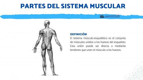
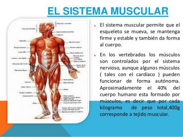
The skeletal muscular system is the active part of the musculoskeletal system. Together with the bones (passive part of the locomotor system) it is responsible for producing movement and giving stability to the body when it is at rest. The skeletal muscular system of the human body is made up of more than 600 muscles and they constitute 40% of the weight of the human body. Most of them make up the musculoskeletal system. In this lesson from a TEACHER we will see what the parts of the muscular system (musculoskeletal system), where they are located and what movements they are responsible for.
Index
- What is the muscular system and what is it for?
- Parts of the muscular system: head and neck
- Trunk muscles
- Parts of the muscular system: muscles of the extremities
What is the muscular system and what is it for?
The system musculoskeletal Is the set of muscles attached to bones of the skeleton. This union can be direct or by means of tendons that join the muscle to the bones. These muscles are made up of
striated muscle tissue, which have the property of being quickly and voluntarily contracted.Voluntary contraction of skeletal muscles is controlled by the brain, (superior structure of the nervous system that is contained within the skull). The voluntary movement the muscular skeletal system requires a certain planning, coordination and precision; which takes place mainly at the level of the motor cortex of the brain.

Parts of the muscular system: head and neck.
We begin by knowing the parts of the muscular system by analyzing the muscles that are in the upper part: in the head and in the neck.
Head muscles
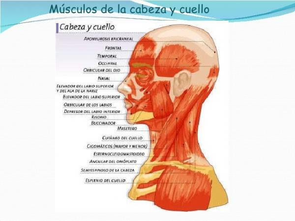
The head contains 30 muscles, which are divided into three large groups based on their origin and the role they perform. They are the chewing muscles and the mimic muscles.
Chewing muscles
They are the ones that are attached to the joint between the skull and the jaw. The function of these muscles is to raise the jaw to bite. The most important are the masseter and the temporal muscle.
Mimic muscles
They are the muscles of the face responsible for facial expressions. They are characterized by being flat muscles that are innervated by the facial nerve. These are flat muscles that have insertion into the skin of the face and neck and they are arranged around the openings of the face (oral cavity, ear cavity, nasal cavity and eye sockets).
The insertion of these muscles to the skin of the face causes the movement of the face when the muscles contract. In addition to generating facial expressions, they are responsible for reflex movements such as palpebral movement (blinking). This is due to the condition of mixed muscles of the palpebral muscles that allow both voluntary and involuntary movement (reflex).
An example of mimic muscle is the giggly, which is involved in the formation of the smile. This muscle is inserted in the corner of the lips and causes, when contracting, the retraction of the labial commissure (increases its transverse diameter).
The calvaria muscles They are the muscles that cover the upper part of the dome of the skull. They are connected to the scalp and are flat and thin, their contraction causes the elevation of the eyebrows and the formation of wrinkles on the forehead so they can also be considered muscles mimics.
Neck muscles
All the neck muscles are mostly long and thin. The muscles of the deeper areas are involved in stabilizing the head while the superficial muscles of the the nape and the lateral area of the neck allow a wide range of head movements and also allow flexion of the neck. In addition, some of those located in the deepest area are involved in the process of swallowing food (swallowing).
One of the most important muscles in the neck is the muscle sternocleidomastoid, on the side of the neck. It is a long and robust muscle that is involved in flexion, rotation and tilt movements of the head.

Trunk muscles.
We continue to know the parts of the muscular system to speak, now, of the muscle groups that are in the trunk.
Posterior or dorsal muscles
The back muscles form several overlapping layers that protect the spine. Acting at the same time these muscles support and keep the spine erect. If only the muscles on one side of the back act, there is rotation and / or flexion of the back towards that side.
Abdominal and thoracic muscles
They are the muscles of the anterior (front) part of the trunk. The thoracic muscles are involved in the adduction movement (approaching the arm to the trunk) the forelimbs (arms) as well as in flexion movements of the trunk.
The muscles of the abdomen have multiple functions because, in addition to allowing the movement of the trunk, they protect and keep in place the different organs that the abdominal cavity contains. It also participates in different physiological processes because its contraction increases the intra-abdominal pressure that facilitates defecation, urination or childbirth, in addition to facilitating inspiration and expiration in the breathing.
Deep
The deep muscles of the trunk have a stabilizing function of the spine. They are innervated by vertebral nerves.

Parts of the muscular system: muscles of the extremities.
We will now know the muscles of the extremities in order to know how the muscular system is formed.
Forelimbs or upper extremities
The arm muscles are divided into 4 groups:
- The shoulder muscles: that allow lifting and moving the arms.
- The arm muscles: In the extremities we find two groups of muscles that are respectively, antagonistic muscles. The antagonist muscles are those that perform contrary actions to carry out a movement. That is, during movement one of the muscles (agonist) contracts to produce the movement of the limb, while the opposing muscles that relax to allow said movement. In the case of the arm, in the flexion movement, the main agonist muscle is the biceps contracting, while the main antagonist muscle is the triceps which relaxes to allow flexion of the elbow.
- The forearm muscles: which are those that allow the rotation of the forearm and the movement of the hand in any direction and flexion and extension of the fingers. It is a very large group of muscles.
- The muscles of the hand: these are muscles that allow the movement of the fingers independently. They are short muscles of small size, since they originate and insert in the bones of the hand.
Hind or lower extremities
They have muscle groups equivalent to those of the forelimbs. Thus we distinguish 4 groups.
- Hip muscles: These are the muscles that make up the buttocks (glutes) and are involved in maintaining an upright posture and play an important role in bipedal locomotion.
- Thigh muscles, as in the case of those on the arm, are antagonistic muscles. The most important are the quadriceps, which allows leg extension; and his antagonist, the triceps femoris. In addition, in the thigh are also the abductor muscles that allow the extension and flexion of the thigh.
- Leg muscles: the set of leg muscles allow flexion of the foot in bipedal locomotion, the most important are soleus and twins that are inserted into the calcaneus bone of the foot through the Achilles tendon.

If you want to read more articles similar to Parts of the muscular system, we recommend that you enter our category of biology.
Bibliography
Netter, Frank H. (2019). Atlas of Human Anatomy - 7th Edition Barcelona: Elsevier España, S.L.U.

