Function of MUSCLES and muscle tissue

The muscles are the anatomical structures constituted by muscle tissue. There are different types of muscle tissue and each of them performs a specific function. In this lesson from a TEACHER we will see what are the different types of muscle tissue and the muscle function.
Index
- The muscles of the body and muscle tissue
- Types of muscle tissues
- Function of skeletal, skeletal, or voluntary muscle
- Functions of cardiac or myocardial muscle tissue
- Smooth muscle tissue functions
The muscles of the body and muscle tissue.
Before knowing what the function of muscles is, let's talk about muscle tissue that is characterized by three fundamental properties: These organs are made up of muscle tissue that is made up of cells called myocytes or contractile fibers.
The shrinkage capacity of muscle fibers is due to the presence of two proteins in this type of cells: actin and myosin. These are proteins that form microfilaments. During muscle contraction, actin binds to myosin.
The binding of actin and myosin fibers it causes the conformation change of myosin that folds in an articulated way. The shortening of the myosin pulls the actin bound to it and results in the myosin being slide on filamentous actin molecules causing shortening of the muscle fiber (contraction).
Different muscle tissues have a closely characteristic morphology and organization of muscle fibers. related to the type of function they perform.
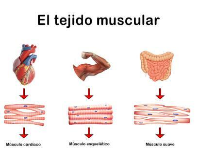
Types of muscle tissues.
To know better what the function of the muscles is, we must first know what are the different types of muscle tissues. Here you have them:
Striated or skeletal muscle tissue
Striated muscle tissue is made up of elongated, plurinucleated cells (with several nuclei). These cells are called myocytes. Inside the myofibrils (actin and myosin fibers are arranged to form transverse striae.
The contraction of these cells is quick and voluntary (with the exception of the diaphragm muscle, whose movement is not voluntary).
Three types of skeletal muscle are distinguished:
- Red fibers or type I fibers: they are small muscle cells, with a large number of mitochondria and highly vascular (supplied by a large number of blood vessels). They are responsible for movements that require little energy and are prolonged, such as maintaining posture. They are muscle fibers resistant to fatigue.
- White or type II fibers: They are muscle cells with a diameter greater than the red fibers. They have few mitochondria and are poorly vascularized. On the other hand, they have a lot of glycogen (energy reserve polysaccharide). They are very powerful fibers, but little resistant to fatigue and are involved in brief but intense exercises.
- Type IIa fibers: They are muscle cells intermediate between type I and type II fibers. Depending on the exercise, they can evolve into type I fibers, depending on the type of exercise and training performed. Strength training, with prolonged and moderate exercises. If the type of training is resistance with short exercises (between 30 seconds and 2 minutes) and intense.
Striated muscle tissue of the heart or myocardium
Cardiac muscle tissue is made up of two different cell types: myocardial cells and cells of the cardionector or autonomic system. The contraction of myocardial cells is rhythmic and continuous.
Myocardial muscle cells are cells with a single nucleus and with striated myofibrils (with layers of actin and myosin arranged in layers). These cells have lateral processes that put them in contact with the processes of other cells. myocardial, the contact between processes occurs through specialized structures called discs intercalary.
In addition to myocardial cells, cardiac tissue is also made up of cells of the autonomic or cardionector system. They are modified muscle cells, capable of transmitting nerve impulses. These cells branch throughout the heart forming the so-called Purkinje system. They are the pacemaker of the heart, responsible for the coordination of the myocardial cells that give rise to the cardiac movements of systole and diastole. They are Rhythmic and coordinated involuntary contractions.
Smooth, involuntary, or flat muscle tissue
Formed by spindle cells, with a single nucleus and that present myofibrils that are not organized into striae. They are cells that, when observed under the microscope, do not present transverse streaks but rather have a smooth appearance. Its speed of contraction is between 10 and 100 times slower than that of skeletal muscle. His contraction is slow and involuntary with the exception of the urinary bladder muscles.
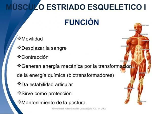
Striated, skeletal, or voluntary muscle function.
We have already seen that, depending on the type of muscle, the functions they provide us can be one or the other. Here we indicate what are the functions of skeletal, skeletal or voluntary muscle
The skeletal muscle is the majority component of the mass of Vertebrate animals. These are muscles attached to the bones of the skeleton, to tendons associated with these or attached to other tissues such as the oculomotor muscles attached to the eyeball.
- Execution ofvoluntary movements: It is one of the main functions of skeletal muscle. Voluntary movements can be defined as those movements that are directed towards a goal, that are consciously controlled and that can be modified at will by the individual. Examples of voluntary movements are locomotion, active touch, eye movements, manipulation of objects, chewing, etc.
- Proprioception: It is the ability to perceive the movements and posture of one's own body regardless of vision. The fact that skeletal muscle works closely with nerve endings that send and receive information to the brain gives rise to skeletal muscle being the main source of information about the body's own disposition in the space.
- Thermoregulation: Skeletal muscle is involved in the thermoregulatory response to cold, especially when other mechanisms fail to compensate for the drop in body temperature. In this case, the skeletal muscle cells increase their activity by means of serial contractions and repetitive (tremors), releasing residual heat that contributes to the maintenance of body temperature in cold environments.
Functions of cardiac or myocardial muscle tissue.
The heart muscle it has structural and functional characteristics intermediate between skeletal muscle and smooth muscle. The function of these muscles is to provide the heart with a rhythmic and continuous contraction, that allows the blood pumping.
These rhythmic contractions are possible thanks to the existence of the cardiac conduction system formed by the cardiac nodes (the atrial node or sinus node and the atrioventricular node), the bundle of His (strands of modified muscle cells that divide into two branches) and Purkinje cells.
Both in the atria and ventricles have structures called cardiac nodes where the nerve impulse is generated and transmitted. the Hiss beam (.the one that causes the ventricles to contract. This is possible thanks to the presence of the cells that form the Purkinje system that are responsible for generating and transmit the electrical impulse to the heart's ventricles at high speed, causing them to contract during systole cardiac.
The heartbeat is generated in the cells of the sinus node (which is considered the natural pacemaker of the heart) located at the entrance to the atria. This impulse generated in the sinus node is transmitted as a wave from top to bottom along the two atria causing the contraction that drives the blood from the atria to the ventricles.
After traveling along the atria, the electrical impulse reaches the atrioventricular node, which is located between the atria and the ventricles. This node receives the electrical impulse and transmits them to the walls of the ventricles through the right and left branches of the Bundle of His, so that the ventricles contract, expelling blood from the heart. At the same time, the atria relax allowing the entry of blood from the veins and preparing for a new contraction, starting the cycle again.

Smooth muscle tissue functions.
We end this lesson with the function of muscles by talking about those that are smooth muscles. The smooth muscle It covers all the internal cavities of the hollow organs of the body with the exception of the heart. Smooth muscle function will vary depending on the type of organ it is in.
- Digestive system: the smooth muscle of the digestive tract has the function of advancing its content. Contraction of the smooth muscle of the intestinal tract is known as peristalsis. It is a progressive contraction from one end of the intestinal tube to the other that causes the ingested food to advance.
- Blood vessels: the smooth muscle that lines the interior (tunica media) is more developed in the arteries than in the veins. It performs two essential functions: Its function is keep blood pressure constant despite changes in blood flow and blood pressure caused by cyclical movements of the heart. Regulate blood flow in the different parts of the body depending on the needs of each moment.
- Uterus: The smooth muscle tissue of the uterus behaves differently during pregnancy and menstruation. Hormones and nerve connections are capable of modifying even the characteristics of muscle tissue. During pregnancy and childbirth, there are contractions of the smooth muscle of the uterus that increase in frequency and intensity.
- Urinary bladder: Contractions of the smooth muscle of the bladder occur when it fills with urine, which causes the emptying of this in the process of urination. Urination is a voluntary process that begins when the volume of the bladder reaches a certain value (generally when they have been stored in the bladder about 200 ml of urine), at this time, the brain receives a signal that the bladder is full and the person feels the need to to pee. A baby's bladder contracts when the bladder reaches a certain volume. As the child grows, her brain learns to control the contraction of the bladder muscle so that it can voluntarily delay urination. It is therefore the only case in which smooth muscle contracts voluntarily.

If you want to read more articles similar to Muscle function, we recommend that you enter our category of biology.
Bibliography
Hershel Raff, Michael Levitzky (2013). Medical physiology. An apparatus and systems approach. Madrid: McGraw-Hill Interamericana de España S.L.