The different PARTS of a VERTEBRA

Image: Natural Sciences
The vertebrae are the bones that make up the spine of human beings. The spinal column is a very large bony organ that runs through many parts of our body, and therefore in it we can find different areas or regions, with specialized functions. While the vertebrae of the cervical region have a lot of mobility, those of the lumbar region have less mobility and their function is basically to support the weight of the upper part of the trunk. Since they have different functions and characteristics, the parts of a vertebra they will be different depending on the type of vertebra we are talking about. In this lesson from a TEACHER we talk about this topic.
Index
- Vertebral body, one of the parts of a vertebra
- Vertebral arch
- How is a vertebra articulated?
Vertebral body, one of the parts of a vertebra.
Despite the great differences between the vertebrae of the different parts of the spine, in all of them we can see Two parts very differentiated: the body and the bow which, as we will see below, have different peculiarities.
Vertebral body
It is called body a the anterior part of the vertebras. It usually has a more or less rounded shape and occupies a large part of the vertebra, so the vertebral arch is easily differentiated.
The function of the vertebral body is give resistance to the vertebra, something that we can see in its internal structure. The vertebral body is made up of a cylinder of spongy bone tissue surrounded by a layer of thin cortical bone. This body will be so much bigger in the vertebrae that more weight they have to support.
The lumbar vertebrae, who have to bear the weight of the upper part of our body have a vertebral body much larger and apparently the thoracic vertebrae for example and even larger than those that we can find in the cervical vertebrae, whose vertebral bodies are not very resistant and thin.

Image: Onmeda.es
Vertebral arch.
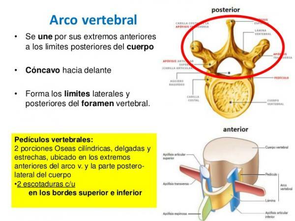
Within the parts of a vertebra we find the vertebral arch which is the back of each of the vertebrae. This part of the vertebra is the one that is attached or anchored to the body, by means of two extensions called pedicles. The pedicles delimit the sides of the foramen of the vertebra.
From the arch we can also find other three types of structures protrusions or extensions, called apophysis or processes. These vertebral projections will have different shape, characteristics and direction depending on the area of the spine that we are in. The vertebral processes of a typical vertebra are:
- Articular processes. The articular processes are attached to the pedicles. In a vertebra we can find four articular processes, a pair in the part that connects the vertebra with the previous one. (superior articular processes) and a pair in the part that connects it with the lower vertebra (articular processes lower).
- Transverse processes. A transverse process is attached to each of these pairs of articular processes, therefore, in each vertebra we will have a total of two transverse processes. The transverse processes can be easily distinguished as they are the only two lateral extensions that the vertebra has.
- Spinous apophysis. These vertebral projections are located on the back of the vertebra, on the opposite side of the body. In each of the vertebrae there is only one spinous process, which has two very important functions: it is responsible for protecting by in front of the medullary canal (which guards the spinal cord) and serves as a place of attachment or insertion with the powerful muscles of the trunk.
Uniting the transverse processes and the spinous process we find the vertebral laminae. The vertebral laminae are found in the postero-lateral part of each vertebra. It is shaped like a semicircle and, thanks to its continuity with the posterior part of the body of the vertebra, forms a closed circle called spinal foramen. The spinal foramen of all vertebrae forms a tube, through which the spinal cord passes, called medullary canal.

Image: Slideshare
How is a vertebra articulated?
Each of the vertebrae in our body has three points of articulation.
- Intervertebral discs. On the one hand, the vertebral bodies are related to each other through the intervertebral discs. The intervertebral discs produce a flexible yet strong joint and also cushion the compressive forces to which the spine is subjected. In anatomy, this joint is classified as a cartilaginous joint or amphiarthrosis.
- Zygapophyseal joint. On the other hand, in each vertebra we find a zygapophyseal joint or interpophyseal joint. This type of joint is of the synovial type and is the one that occurs between the superior articular process of a vertebra and the lower articular process of the vertebra that is directly about her. This type of joint is really in charge of driving and limiting the movement of the vertebrae between them, so They are usually the most affected by degeneration and wear and tear typical of age or by dislocations, fractures and processes of osteoarthitis.
- Spinal hole. Finally, the vertebrae are also joined together by their central part, the spinal foramen. As mentioned above, these joined holes form the spinal canal or medullary canal, through which the spinal cord passes. Between vertebra and vertebra we can also find an intervertebral foramen, an exit conduit for the vertebral nerves.
Together we can say that a typical vertebra has: a body and an arch, the latter consisting of: two vertebral laminae, two pedicles, one spinous process, two transverse processes and four processes articular. It should be clarified that this is not true for the first two vertebrae of the spine, the atlas and the axis. These vertebrae, due to their position and their function, have undergone great variations with respect to this general pattern.
Image source:
If you want to read more articles similar to The parts of a vertebra, we recommend that you enter our category of biology.
Bibliography
- Moore, K. L., & Dalley, A. F. (2009). Clinically oriented anatomy. Panamerican Medical Ed.
- Luque Sendra, María Isabel (2009). Study of the morphology of the vertebral body in a human L4 with models of internal and external bone remodeling (Final Thesis). Higher Technical School of Engineers, Seville, Spain.
- CMT Osteopathy (September 4, 2016). Anatomy of the spine. Recovered from http://www.cmtosteopatia.com/es/articulos/anatom-a-de-la-columna-vertebral, 0.html


