Parts of the eye and their functions

Image: Apanovi
The eye is the most important organ of the eyesight, with which we can capture the images of objects and get information about the shapes, colors, distance, movement or position of these. Next, in this lesson from unPROFESOR.com we will study the parts of the eye and their functions so that you can better understand our human anatomy and how this important organ works in our daily lives.
Index
- Structure of the eye
- The sclera layer of the eye
- Choroid or vascular tunic
- The retina of the eye
- Transparent media
- The eye and the light
Structure of the eye.
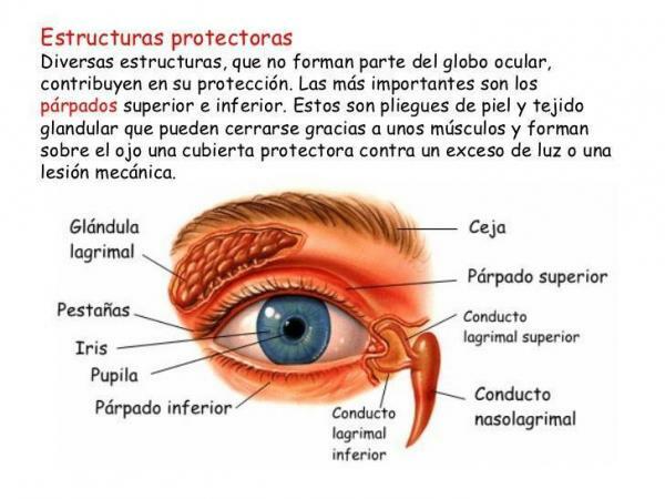
The shape of the eye is spherical and is about 2.5 centimeters in diameter. By the action of six muscles, two oblique and four straight, it moves and is externally protected by the eyelids and eyelashes.
In addition, it is made up of three layerconcentric s:
- The sclera or fibrous tunic
- The choroid or vascular tunic
- And the retina or nervous tunic
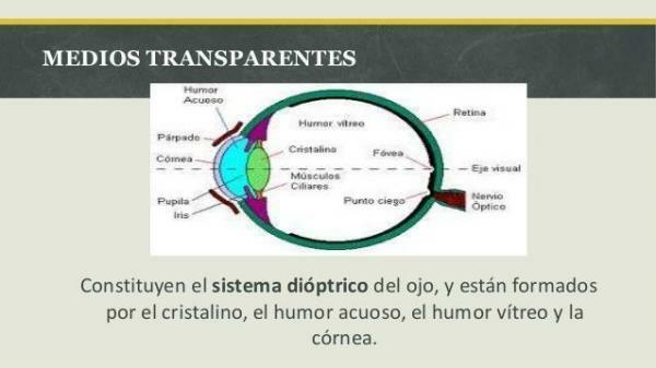
There are also a number of transparent media and refrigerants, among which the cornea, the lens, the aqueous humor and the vitreous humor stand out.

Image: Slideshare
The sclera layer of the eye.
We begin by analyzing parts of the eye and their functions to talk about the sclera layer. It is firm membrane which forms the outer layer of the eye, also known as the fibrous tunic. It is very resistant and is made up of fibrous-connective tissue that preserves the inside of the eye and gives it rigidity.
Its function is protect the sensitive structures of the eye and within it we can distinguish Tenon's capsule, which is a resistant membrane that covers the sclera and that makes up the muscle sheath of the eye, which supports the eye and separates it from the cavity orbital.
Choroid or vascular tunic.
The tunica vascular or media, also known as the choroid, is where the blood vessels are located on which the retina depends. It is located between the sclera and the retina, and covers the inside of the eyeball, whose external face is shiny and black.
In the front, there is a perforation in the center, The pupil, which is surrounded by the iris, circular membrane whose function is to regulate the light that enters through the pupil, contracting or dilating depending on the luminosity. In the posterior area of the choroid is the optic nerve.

Image: Slideshare
The ocular retina.
Within the parts of the eye and their functions we will also talk about the nervous tunica or retina, which is the innermost area of the eye, being an extension of the central nervous system. The optic nerve originates from it and functions as a light-sensitive plate.
Within it, three parts are distinguished:
- The papilla or optic disc: It is the access sector of the optic nerve in the retina, entering the retinal arteries and leaving the retinal veins. It is the blind spot of the eye because it does not have light-sensitive cells.
- Taint: It is located behind the retina and due to the large number of photo-receptors and blood vessels in it, it specializes in fine vision, which is used to perceive details of objects.
- Fovea: it is a small depression that is located in the center of the macula, measuring 1.5 square millimeters, which allows vision of greater precision and sharpness.
Transparent media.
We continue with the parts of the eye and their functions, now analyzing the transparent media, which make up the diopter system, they stand out:
- Cornea. It is located in the anterior area of the sclera and, as a clear and transparent layer, allows light rays to pass through. Due to its regular curvature, it works like a convergent lens, having a radius of curvature of about 8 mm. Therefore, it has two functions: an optical and a protection of the front part of the eye.
- Crystalline. This biconvex lens is located behind the iris and serves to accommodate the eye, that is, to focus precisely. The lens is held in place by the suspensory ligament, also called the zonule of Zinn, which attaches it to the tunica bascular. Small muscles modify its shape and make it more curved to focus on nearby objects and flatten it to see distant objects.
- Aqueous humor. It is a fluid and transparent liquid, 98% made up of water, which is located between the lens and the cornea, in the anterior area of the eye. It keeps the eyeball inflated and oxygenates and nourishes the lens and the cornea, since both have no blood supply.
- Vitreous humor. It is also called the vitreous body or gel, and it is formed by a colorless and gelatinous substance, which fills the back of the eyeball, between the lens and the retina. It maintains the shape of the eye and its internal pressure, and is formed during embryonic life, so it is not renewed. Its function is to maintain the shape of the eyeball and achieve a uniform surface of the retina, so that sharp images can be received.

The eye and the light.
Within the parts of the eye and their functions it is important to describe how light acts on this organ. It enters through the pupil and concentrates on the cornea and lens to form an image on the retina. Keep in mind that the retina has millions of cells, called rods and cones, that are sensitive to light.
Cones require bright light to function and they detect many shades of color. On the other hand, the rods need little light and they do not distinguish the colors very much, being assigned for the vision with darkness.
The nervous excitations that occur in the retina are transmitted by the optic nerves from both eyes in the form of nerve impulses to the cerebral cortex, where the stimuli of visual perceptions and sensations take place. Information from the optic nerves is processed in the brain and results in a coordinated image.
If you want to read more articles similar to Parts of the Eye and Their Functions - For Kids, we recommend that you enter our category of biology.


