ALL BONES of the lower limb
The lower limb bones They can be grouped into four groups, each set of bones has points of union with other bones, the joints. In this lesson from a TEACHER we will see in detail what are the bones of the lower limb and how they are articulated between them.
Index
- Bones of the lower limb
- Pelvic girdle bones
- Thigh bones: femur
- Leg bones
- Foot bones
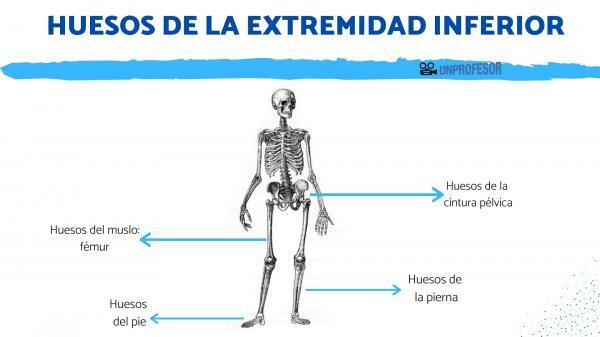
Bones of the lower limb.
This brief diagram shows the components of each of the groups of bones that make up the lower limb. Each of these four groups is developed in more detail in the following sections.
1-. Bones of the pelvic girdle (bony pelvis):
1.1 Iliac or coxal bones
- The ileum
- The ischium
- Pubis
- 1.2 Sacral
- 1.3 Coccyx
2-. Thigh bones
- 1.2 Femur
3-. Leg bones
- 3.1 Patella
- 3.2 Tibia
- 3.3 Fibula
4-. Foot bones
- 4.1 Tarsal bones
- 4.2 Metatarsal
- 4.3 Phalanges
Pelvic girdle bones.
We begin this review of the bones of the lower limb by focusing on the ones we have in the pelvic area.
The set of hip bones is also known as bony pelvis. It is the part of the skeleton that is located in the lower part of the trunk. The bony pelvis is a
musculoskeletal funnel, that tapers down. This bone structure allows bipedal locomotion (to walk upright).During childbirth, the baby must pass through this funnel, for this reason the pelvic girdle is wider in the case of women, this being one of the features of sexual dimorphism that allow the gender of an individual to be identified from its skeletal remains.
1. Iliac or coxal bones
There are two coxal bones and they are located on both sides of the spinal column. They are shaped like an eight and have three distinct regions: ileum, ischium and pubis. These regions correspond to three independent bones in the fetus and distinguishable as units in young individuals; but they are fused in adulthood.
The ileum It is the largest bone of the three that make up the innominate bone, it is shovel-shaped and slightly concave. It forms the top of the innominate bones. The upper border of the ilium is known as iliac crest. The iliac crest is one of the bones that contains the largest amount of bone marrow in adults. The bone marrow It is composed of stem cells that will give rise to the different blood cells and has a fundamental role in the immune system.
The iliac crest is of important clinical interest, since the large amount of bone marrow it contains and the fact that is easily palpable, make the iliac crest one of the most used areas for obtaining marrow that is.
- The ischium: It is located in the lower posterior area of the iliac bone. This bone presents the ischial tuberosity, which is the support support in the sitting position. Another part of this bone is the ischium branch, which is the area where it merges with the pubis, limiting an orifice that is known by the name of obturator foramen of the pelvis. Through the obturator form the blood vessels, the nerves pass outside the pelvis. This overture presents differences in the two genres (sexual dimorphism), the foramen being larger and oval in men and smaller and slightly triangular in shape in women.
- The pubis: it is situated in the lower anterior part of the innominate bone, forming the front part of the pelvic girdle. In the medial area the two pubic bones meet at the joint called symphysis pubis formed by a line of cartilage. The symphysis pubis widens slightly when the legs are wide apart.
- The acetabulum: It is part of the hip joint. The acetabulum is a hemispherical depression (cup-shaped) that forms in the area of contact between the three coxal bones. It is part of the hip joint and articulates with the head of the femur.
2. Sacrum
The sacrum is located at the back of the waist, at the lower end of the spine. It is wedge-shaped and sits between the innominate bones. It is made up of five fused vertebrae. Presents four holes of conjugation through which the spinal nerves (nerves that originate in the spinal cord) of the sacral area.
3. Coccyx
The coccyx is the lower part of the spine, in colloquial language it is known as the tailbone. It is a vestigial tail (remains of a tail that has disappeared throughout the evolutionary process). It is made up of between three and five vertebrae that can be independent or fused. It is a solid bone (or set of bones) in which there is no internal channel through which the bone marrow runs. The variability of this bone is very great, there being a great variability of shapes and sizes between different individuals.
The upper end of the coccyx is attached to the sacrum by means of ligaments.
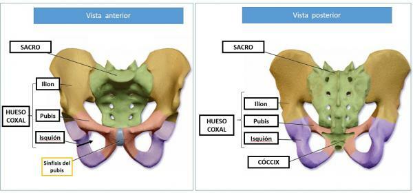
Image: Dolopedia
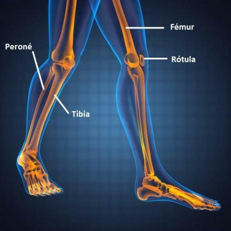
Thigh bones: femur.
The only bone in the thigh is the femur, it is the typical long bone. Is he longest and strongest bone in the skeleton human. It is an asymmetric bone that articulates at its upper end with the hip joint and at its lower end with the tibia. Considering the skeleton in a vertical position, the femur descends obliquely from the outside to the inside and presents a slight twist on its vertical axis.
Both the upper end joint and the lower end joint of the femur have synovial joints. Synovial joints are those in which the articular surfaces of the bones in contact are enclosed in a joint capsule. This capsule is made up of two layers:
- Synovial membrane: an inner layer, which produces the so-called synovial fluid and which has a lubricating function.
- Fibrous membrane: an outermost layer of dense connective tissue that surrounds and stabilizes the joint.
The end or superior epiphysis of the femur, has a spherical shape and receives the name of head of the femur. The head of the femur is the articular surface with which this bone fits into the acetabulum of the coxal or iliac bone, forming the hip joint or pelvic joint.
The epiphysis or lower end of the femur is widened and is shaped like a pulley (trochlea), is divided into two portions of unequal size, the posterior portion being much larger than the anterior. The lateral and medial enlargements of the trochlea are called condilies. The trochlea and the two condyles form the articular surface, which form the knee joint along with the tibia and patella (leg bones).
Leg bones.
We are now going to know the bones of the lower limb that are concentrated in the rest of the leg. The leg bones are as follows.
1. Ball joint
The kneecap or patella is a sesamoid bone, that is, a bone that is attached to a tendon and whose function is to increase the leverage effect of the bone joint on the muscle. This bone is included in the patellar tendon. It is an essential part of the knee joint along with the femur and tibia.
2. Tibia
The tibia is a long, wide, and strong bone that supports the weight of the body; It is located on the anterior and internal face and the larger of the two bones of the leg. The epiphyses (ends of the bone) have two independent joints in both cases.
Its upper epiphysis has a wide and flat surface that is called the tibial plateau. The tibial plateau is the articular surface of the tibia that comes into contact with the femur condyle, in the knee joint. In addition, it also has a posterior lateral articular surface in which it articulates with the fibula.
The lower epiphysis of the tibia forms the internal malleolus (internal bulge of the ankle). This epiphysis on the underside, which has a pulley-shaped surface (trochlea), articulates with the talus (foot bone). In addition to the same way as in the superior epiphysis, it also articulates with the fibula by means of a surface
3. Fibula
The fibula is a long, thin bone that runs parallel to the tibia on the outside of the leg. It practically does not bear weight and several muscles are inserted into it. Its upper epiphysis forms the fibula head which articulates with the tibia below the knee joint.
Its lower epiphysis is longer and wider than that of the tibia and forms the external bulge of the ankle o external malleolus. At the lower end, the fibula also has an articular surface in contact with the talus.
Foot bones.
The foot bones They are also the bones of the lower limb of the body. Here we indicate their names.
1. Tarsal bones
The tarsus is an area of the foot composed of seven bones, which bear most of the body weight. The two largest tarsal bones are the talus and calcaneus. They are located on the back of the foot forming the heel and ankle. The astragalus It is the tarsal bone that is part of the upper ankle joint, known as the head of the talus, where it is in contact with the tibia and fibula. The calcaneus It is the largest of the tarsal bones and forms the heel of the foot.
The five remaining bones that make up the tarsus are smaller and form the midfoot. At their most distal end (end furthest from the central axis of the body) they are in contact with the metatarsal bones.
2. Metatarsus
They are five elongated bones of similar shape but of different size. At its proximal end (closest to the central axis of the body) the metatarsal bones articulate with the tarsal bones. Its distal ends articulate with the bones of the fingers (phalanges).
3. Phalanges
They are a total of 14 short bones that form the toes. Four of the five toes have a total of 3 phalanges while the hallux (big toe) only has two phalanges.

Image: Pinterest
If you want to read more articles similar to Bones of the lower limb, we recommend that you enter our category of biology.
Bibliography
Netter, Frank H. (2019). Atlas of Human Anatomy - 7th Edition Barcelona: Elsevier España, S.L.U.