All BONES of the NECK
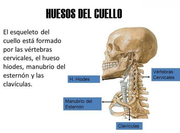
Image: Human skeleton
The neck is the anatomical region that unites the head with the trunk. Inside the neck we can find two types of bones: the hyoid bone, a small and odd bone, with a U-shape that it is located just above the larynx and cervical vertebrae, which form the first region of the spine vertebral. In this lesson from a TEACHER we will review all neck bones, their names, functions and characteristics main. If you want to know more about the bones that make up your neck, keep reading!
Index
- The neck and its bones
- The hyoid bone
- The cervical vertebrae: main characteristics
The neck and its bones.
The neck is a fairly complex junction structure, which allows great movement capacity (flexion, extension and rotation movements of different kinds) but it also helps to support and protect the spinal cord that contains the spinal column. The spinal cord is especially delicate in this region of the body as it is more exposed than in areas lower back where, in addition to the spine, large muscles, fat deposits, etc. that help to protect you.
The only bone that is properly the neck is called hyoid and it is located just above the larynx, at the top of the neck. In addition to the hyoid bone, in this lesson from a TEACHER we will discuss the cervical vertebrae They can be classified into three groups of bones: neck bone, spinal bone or chest bone.

Image: Slideshare
The hyoid bone.
Within the bones of the neck, the hyoid is the one that is mainly found in this region. It's a small, odd bone, which is located in the upper part of the neck, above the larynx and at the approximate height of the third cervical vertebra. This bone is U-shaped, with the greater horns (branches of the U) towards the nape of the neck and the body (central part or "belly" of the U) towards the front of the neck (ventral).
The hyoid bone it's a free bone, which does not articulate directly with any other, but is suspended in position. However, this bone is related (via ligaments and other structures) to three regions of the neck and head:
- In the upper part it is attached to the floor of the oral cavity.
- At the bottom, the hyoid is attached to the larynx.
- At the back or back of the neck, the hyoid is attached to the pharynx.
This location makes the hyoid a bone that, despite being very mobile, is very strong and is capable of serving as a bone anchor to many structures and muscles with which it is related. In addition, the hyoid bone is highly protected so injuries to it are not very common. A hyoid injury or tear is indicative of severe trauma (significantly affecting the ability to speak and breathe) or strangulation.

Image: Slideshare
The cervical vertebrae: main characteristics.
Another of the neck bones are this type of vertebrae. Humans have seven cervical vertebrae (C1-C7) that are notably smaller than the rest of the vertebrae, they have a foramen in each of the transverse processes, the spinal bifid process, etc. Also, the first two cervical vertebrae have some features that differentiate them from the rest:
- C1 or atlas. His name comes from the Greek titan condemned by Zeus to carry the sky on his shoulders since the first vertebra cervical supports the weight of the skull, which is directly resting on this vertebra as Atlas carries the Darling. This vertebra lacks a vertebral body and spinous process, which are replaced by two lateral masses that are connected by an anterior and posterior arch. In addition, this vertebra fuses with the second cervical vertebra to support weight more effectively.
- C2 or axis. This vertebra also contains bilateral masses, which allow it to articulate with C1; This joint allows the Atlas to rotate on the axis and thus the rotational movement of the neck and head can be produced. In addition, it also has an odontoid process, or “dens”, in the upper aspect of the body that allows it to better articulate with the C1.
- C3 to C7. The cervical vertebrae that follow the atlas and axis are quite the same between them and with respect to the rest of the vertebrae that make up the spinal column. In addition to giving mobility to the neck and head, they allow their extension and contraction.
Injuries to the cervical vertebrae
While injuries to the hyoid bone are quite rare, injuries to the cervical vertebrae are quite common and some of them very serious. The two best known injuries are:
- Whiplash or hyperextension injury. It occurs when the head is struck back on the shoulders more or less static, causing a whiplash. Whiplash is very common in traffic accidents in which there is a collision from behind, but it can also occur while playing rugby or certain types of falls. In most cases, what occurs is an injury to the anterior longitudinal ligament of the cervical vertebrae, but in other more serious cases, fractures can also occur in the cervical vertebrae. own vertebrae (which undergo strong compression movements) or dislocations of the C2 on the C3, which can even damage the spinal cord and result in tetraplegia or death.
- Jefferson fracture of the atlas. It occurs when there is a vertical drop on an extended neck, for example when diving in excessively shallow water. This fall compresses the lateral masses of the Atlas between the occipital condyles and the axis, which separate, fracturing one or both anterior / posterior arches. In other more serious cases, a tear or injury of the transverse Atlas ligament may occur.
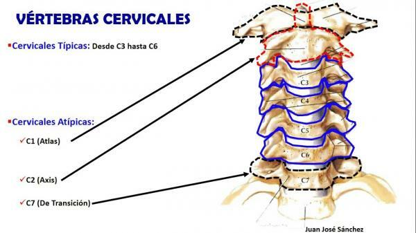
Image: Youtube
If you want to read more articles similar to All the bones of the neck, we recommend that you enter our category of biology.
Bibliography
- Waxenbaum, J. A., & Futterman, B. (2018). Anatomy, back, cervical vertebrae. In StatPearls [Internet]. StatPearls Publishing. Recovered from https://www.ncbi.nlm.nih.gov/books/NBK459200/
- Schmidler, C. (April 23, 2018). Neck Anatomy Pictures Bones, Muscles, Nerves. Recovered from https://www.healthpages.org/health-a-z/neck-anatomy-pictures-bones-muscles-nerves/
- Monasterio, A. (January 16, 2017). Hyoid bone. Recovered from https://www.blogdefisioterapia.com/hueso-hioides/
- Topographic anatomy. (s.f). Cervical vertebrae. Recovered from https://www.anatomiatopografica.com/huesos/vertebras-cervicales/
- Topographic anatomy (s.f). Hyoid bone. Recovered from https://www.anatomiatopografica.com/huesos/hueso-hioides/