Anatomy of the vertebral column

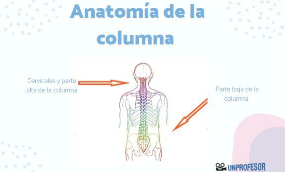
The spine is one of the largest systems in the body. Made up of bones, muscles and cartilage, it forms a group capable of supporting and protecting the human body, at the same time that it gives the necessary flexibility to curve our body, walk and perform a large amount of movements.
In this lesson from a TEACHER we will review spine anatomy and we will answer frequent questions such as what the spine is, what it does and what its parts are. If you want to know more, we invite you to continue reading!
Index
- Definition of the spine
- The vertebrae and intervertebral discs
- Ligaments and muscles of the spine
Definition of the spine.
To know the anatomy of thespine we must bear in mind that it is the basic structure of the body. It is defined as a osteofibrocartilaginous system, articulated and resistant. This is because it is formed, in the adult human being, by:
- 50 bones
- 120 joints
- 23 intervertebral discs
- 33 vertebrae, divided into 5 zones
Functions of the spine
This complex system is interconnected to give rise to a longitudinal axis or stem, that completely runs through the posterior and lower portion of the human skeleton. Its position in the human being has been very important in its evolution, since it has the following functions:
- Offers support for to the head, upper limbs and rib cage during movements and weight-bearing activities.
- Toast protection to vital organs (such as the heart and lungs), but also to soft tissues, such as the spinal cord spinal cord, during physiological movements and load-bearing activities weight.
- It is a structure for the insertion of muscles of the abdomen and thorax, as well as for some muscles of the upper and lower limbs. All of them are strong muscles, which require a structure like the spine to support their movement.
- Allows the movement throughout its length and power the movements of the upper and lower extremities.
- Increase the visual and auditory fields. Standing led to adaptation of the spine, which led to raising the position of the head with respect to the ground.
- The spinal column allows to maintain the human body both in the static postures as in the dynamics, facilitating the passage from the first to the second.
- Finally, the spine acts as a device for shock absorption during travel.
The vertebrae and intervertebral discs.
We continue to know the anatomy of the spine to focus on its main parts: the vertebrae and intervertebral discs.
Vertebrae
The vertebraeThey have different shapes and characteristics, depending on the area or height where they are. Mainly, the spine is divided into five regions:
- cervical region: formed by the first seven vertebrae, C1-C7.
- dorsal or thoracic region: made up of 12 vertebrae, T1-T12
- lumbar region: composed of 5 vertebrae, L1-L5.
- sacro-axial region: also composed of 5 vertebrae, S1-S5.
- coccyx: formed by 4 vertebrae, welded together in adults.
Intervertebral discs
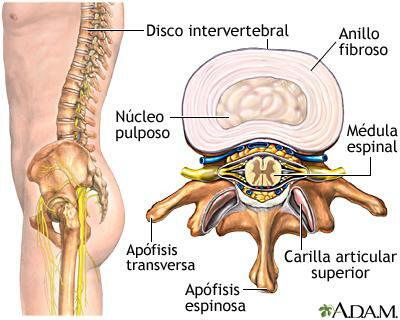
The intervertebral discs they are found at the junction of two vertebrae. Each intervertebral disc consists of a strong fibrous annulus on the outside and a soft gelatinous nucleus, called the nucleus pulposus, on the inside. This allows it to be a viscoelastic structure, that is, it works as a compact mattress that facilitate and restrict the movements that take place in the region. where the vertebral bodies are: in addition, they are capable of transmitting the load from one vertebral body to the next, allowing for example that we let's move.
The intervertebral discs account for approximately 20% - 30% of the height of the healthy spine. If the nucleus pulposus is lost, as occurs with aging, the space between the vertebra and vertebra is reduced. This can cause an increase in back pain and even vertebral subsidence and an increase in dorsal kyphosis.

Image: Medline Plus
Ligaments and muscles of the spine.
The vertebrae are not isolated bones, but are found attached to ligaments and muscles adjacent.
Ligaments of the spine
On the one hand, the vertebrae are attached to the following ligaments:
- The vertebral bodies are attached to the coccyx by the anterior longitudinal ligament (ALL) in front and by the posterior longitudinal ligament (LLP) behind.
- The vertebral arches are joined by the yellow ligaments, which have that color due to the greater amount of elastin, which allows them to flex and stretch the spine.
- The processes are joined by the intertransverse ligaments and interspinous ligaments.
- The supraspinatus ligament that jumps from one spinous to another. It is the most superficial ligament of all and in fact it can be seen when we arch our back.
Spinal muscles
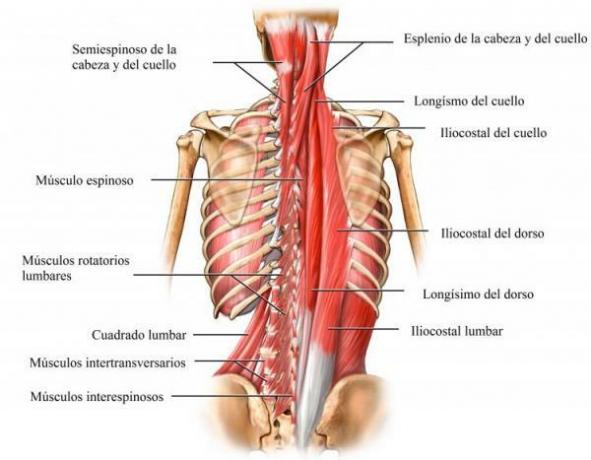
The spinal column, through its numerous bony processes, offers direct or indirect areas of insertion to a series of muscles called axial musculature. The axial musculature is the autochthonous or proper muscles of the body. These muscles are responsible for: stabilizing each of the segments or parts of the spine during movement and normal posture; allow the production of gross movements in many of them and stabilize and produce the physiological movements of the limbs in relation to the trunk. Some muscles closely related to the anatomy of the spine are:
- Serratus posterior, superior and inferior.
- Erect muscle of the spine, formed pro iliocostal, longissimo and spinous.
- Spinotransverse of the head and neck.
- Transversospinosus: formed by semispinosos, multifidos and rotators.
- Suboccipital muscles.
- Hemispinal muscle of the head

Image: City Sports
If you want to read more articles similar to Spinal anatomy, we recommend that you enter our category of biology.
Bibliography
- Luque, S., & María, I. (2009). Study of the morphology of the vertebral body in a human L4 with models of internal and external bone remodeling. Esc. Super technique. Ing. Seville, 30-37.
- Therapeutic Medical Center and Osteoarticular Diseases (September 4, 2016). Anatomy of the spine. Recovered from: http://www.cmtosteopatia.com/es/articulos/anatom-a-de-la-columna-vertebral, 0.html
- Wikipedia (July 3, 2020). Spine. Recovered from: https://es.wikipedia.org/wiki/Columna_vertebral.


